Dear MRI Insider,
I wanted to let you know about two brand new MRI stories we've posted today.
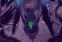
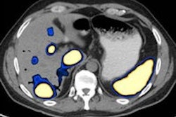
The first is about the added value of diffusion-weighted imaging in suspected cases of rectal cancer. At the 2013 European Society of Gastrointestinal and Abdominal Radiology congress, U.K. authors spelt out exactly why the technique has become an important complement to other sequences in oncology imaging. Get the story here.
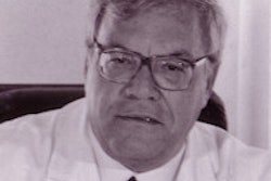
The second article is about MRI pioneer Dr. Roberto Passariello, who died on 11 August at the age of 72. As well as being a past president of the ECR and European Association of Radiology, Passariello used to preside over the European Society of MR in Medicine and Biology, and his department installed the first extremity MRI system in Italy. Click here to read more about his life and career.
In spite of Europe's economic difficulties, the future prospects for MRI seem relatively bright. Our review of the latest market trends focuses on three key areas of potential growth: neurology, extremity scanning, and PET/MRI. For the full details, click here.
At the London Olympics, nearly half of the 1,711 radiological investigations conducted in the polyclinic involved MRI, and the modality proved particularly useful for imaging of elbow pain. Our news report contains details of some interesting cases from the games. Find out more here.
The European Society of Cardiology's annual meeting begins soon in Amsterdam, and MRI looks sure to feature in the imaging sessions at this major event. We'll be covering the meeting, and to limber up for it, we've produced a report about MRI and diastolic function. To view our article, click here.
MRI is developing a central role in dementia. Researchers in Geneva have published an authoritative overview article about this topic in European Radiology, and you can read more here.
This is a small selection of the articles posted in the MRI Digital Community. Please do check out the rest of them below this message.


![Overview of the study design. (A) The fully automated deep learning framework was developed to estimate body composition (BC) (defined as subcutaneous adipose tissue [SAT] in liters; visceral adipose tissue [VAT] in liters; skeletal muscle [SM] in liters; SM fat fraction [SMFF] as a percentage; and intramuscular adipose tissue [IMAT] in deciliters) from MRI. The fully automated framework comprised one model (model 1) to quantify different BC measures (SAT, VAT, SM, SMFF, and IMAT) as three-dimensional (3D) measures from whole-body MRI scans. The second model (model 2) was trained to identify standardized anatomic landmarks along the craniocaudal body axis (z coordinate field), which allowed for subdividing the whole-body measures into different subregions typically examined on clinical routine MRI scans (chest, abdomen, and pelvis). (B) BC was quantified from whole-body MRI in over 66,000 individuals from two large population-based cohort studies, the UK Biobank (UKB) (36,317 individuals) and the German National Cohort (NAKO) (30,291 individuals). Bar graphs show age distribution by sex and cohort. BMI = body mass index. (C) After the performance assessment of the fully automated framework, the change in BC measures, distributions, and profiles across age decades were investigated. Age-, sex-, and height-adjusted body composition reference curves were calculated and made publicly available in a web-based z-score calculator (https://circ-ml.github.io).](https://img.auntminnieeurope.com/mindful/smg/workspaces/default/uploads/2026/05/body-comp.XgAjTfPj1W.jpg?auto=format%2Ccompress&fit=crop&h=100&q=70&w=100)


![Overview of the study design. (A) The fully automated deep learning framework was developed to estimate body composition (BC) (defined as subcutaneous adipose tissue [SAT] in liters; visceral adipose tissue [VAT] in liters; skeletal muscle [SM] in liters; SM fat fraction [SMFF] as a percentage; and intramuscular adipose tissue [IMAT] in deciliters) from MRI. The fully automated framework comprised one model (model 1) to quantify different BC measures (SAT, VAT, SM, SMFF, and IMAT) as three-dimensional (3D) measures from whole-body MRI scans. The second model (model 2) was trained to identify standardized anatomic landmarks along the craniocaudal body axis (z coordinate field), which allowed for subdividing the whole-body measures into different subregions typically examined on clinical routine MRI scans (chest, abdomen, and pelvis). (B) BC was quantified from whole-body MRI in over 66,000 individuals from two large population-based cohort studies, the UK Biobank (UKB) (36,317 individuals) and the German National Cohort (NAKO) (30,291 individuals). Bar graphs show age distribution by sex and cohort. BMI = body mass index. (C) After the performance assessment of the fully automated framework, the change in BC measures, distributions, and profiles across age decades were investigated. Age-, sex-, and height-adjusted body composition reference curves were calculated and made publicly available in a web-based z-score calculator (https://circ-ml.github.io).](https://img.auntminnieeurope.com/mindful/smg/workspaces/default/uploads/2026/05/body-comp.XgAjTfPj1W.jpg?auto=format%2Ccompress&fit=crop&h=112&q=70&w=112)


