
Dear MRI Insider,
Men are living longer and healthier lives, but a downside is that many more of them are being diagnosed with prostate cancer. The disease is now reaching epidemic proportions and this has focused attention on the potential value of prostate MRI, particularly given the modality's ability to demonstrate functional anatomy.
MRI offers great promise in terms of earlier tumor detection, improvement of tumor localization, and better detection and determination of cancer aggression.
But the widespread lack of standardization and structured reporting in prostate MRI remains a key issue, so the European Society of Urogenital Radiology's latest guidance is timely and practical. This is today's top story on AuntMinnieEurope.com, and you can visit our MRI Digital Community to find out more.
Other important publications have taken place during the past month. For instance, researchers have shown that susceptibility-weighted MRI (SWI) is a viable alternative to perfusion-weighted MRI to assess reduced blood flow and predict stroke evolution, according to a study published online on 10 February in European Radiology. The major benefit of SWI in stroke cases is that the technique does not require a contrast agent. Get the story here.
In a new multiple sclerosis study, a German team used 7-tesla MRI with a T1-weighted magnetization-prepared rapid acquisition and multiple gradient-echo (MP-RAGE) technique, and found that the protocol clearly delineated cortical lesions. To learn more, click here.
Heart failure is associated with gray-matter loss at MRI and a worsening of both memory and psychomotor skills. To a lesser extent, the presence of ischemic heart disease slows recall as well, according to a new study in the European Heart Journal. Find out more here.
It appears the Mediterranean diet may well have significant benefits for the brain. New research may help explain why it has been linked to lower incidence of neurological conditions such as mild cognitive impairment, Alzheimer's disease, and dementia. Click here for our article.
The European Congress of Radiology (ECR) begins next week, and MRI will feature prominently in our news reports. Log on starting on Thursday 1 March for our live coverage from Vienna.

![Overview of the study design. (A) The fully automated deep learning framework was developed to estimate body composition (BC) (defined as subcutaneous adipose tissue [SAT] in liters; visceral adipose tissue [VAT] in liters; skeletal muscle [SM] in liters; SM fat fraction [SMFF] as a percentage; and intramuscular adipose tissue [IMAT] in deciliters) from MRI. The fully automated framework comprised one model (model 1) to quantify different BC measures (SAT, VAT, SM, SMFF, and IMAT) as three-dimensional (3D) measures from whole-body MRI scans. The second model (model 2) was trained to identify standardized anatomic landmarks along the craniocaudal body axis (z coordinate field), which allowed for subdividing the whole-body measures into different subregions typically examined on clinical routine MRI scans (chest, abdomen, and pelvis). (B) BC was quantified from whole-body MRI in over 66,000 individuals from two large population-based cohort studies, the UK Biobank (UKB) (36,317 individuals) and the German National Cohort (NAKO) (30,291 individuals). Bar graphs show age distribution by sex and cohort. BMI = body mass index. (C) After the performance assessment of the fully automated framework, the change in BC measures, distributions, and profiles across age decades were investigated. Age-, sex-, and height-adjusted body composition reference curves were calculated and made publicly available in a web-based z-score calculator (https://circ-ml.github.io).](https://img.auntminnieeurope.com/mindful/smg/workspaces/default/uploads/2026/05/body-comp.XgAjTfPj1W.jpg?auto=format%2Ccompress&fit=crop&h=100&q=70&w=100)
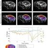


![Overview of the study design. (A) The fully automated deep learning framework was developed to estimate body composition (BC) (defined as subcutaneous adipose tissue [SAT] in liters; visceral adipose tissue [VAT] in liters; skeletal muscle [SM] in liters; SM fat fraction [SMFF] as a percentage; and intramuscular adipose tissue [IMAT] in deciliters) from MRI. The fully automated framework comprised one model (model 1) to quantify different BC measures (SAT, VAT, SM, SMFF, and IMAT) as three-dimensional (3D) measures from whole-body MRI scans. The second model (model 2) was trained to identify standardized anatomic landmarks along the craniocaudal body axis (z coordinate field), which allowed for subdividing the whole-body measures into different subregions typically examined on clinical routine MRI scans (chest, abdomen, and pelvis). (B) BC was quantified from whole-body MRI in over 66,000 individuals from two large population-based cohort studies, the UK Biobank (UKB) (36,317 individuals) and the German National Cohort (NAKO) (30,291 individuals). Bar graphs show age distribution by sex and cohort. BMI = body mass index. (C) After the performance assessment of the fully automated framework, the change in BC measures, distributions, and profiles across age decades were investigated. Age-, sex-, and height-adjusted body composition reference curves were calculated and made publicly available in a web-based z-score calculator (https://circ-ml.github.io).](https://img.auntminnieeurope.com/mindful/smg/workspaces/default/uploads/2026/05/body-comp.XgAjTfPj1W.jpg?auto=format%2Ccompress&dpr=2&fit=crop&h=167&q=70&w=250)


