Positron emission mammography (PEM) is proving a useful technique in the presurgical assessment of invasive lobular carcinoma (ILC), according to research presented at the European Society of Breast Imaging (EUSOBI) annual meeting, held recently in Aberdeen, U.K. Its ability to exclude bilateral disease can also be beneficial.
PEM is a high-resolution tomographic method for molecular imaging of positron-emitting isotopes. It uses a pair of dedicated gamma radiation detectors placed above and below the breast and mild breast compression to detect coincident gamma rays after administration of F-18 FDG.
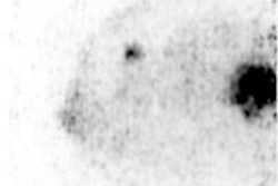
 PEM detected contralateral malignancy missed in conventional mammography. Notably, there is a quantitative difference in uptake between both lesions, with the uptake of IDC in the right breast higher than ILC in the left breast. All figures courtesy of Dr. Aya Deabes et al and presented at EUSOBI 2025.
Dr. Aya Deabes, a radiologist at National Cancer Institute Cairo University, Baheya Foundation for Breast Cancer in Cairo, and Oxford University Hospitals NHS Foundation Trust, U.K., and colleagues conducted a retrospective review of all PEM studies in 2022 and 2023. They identified 46 patients with histopathologically proven ILC.
ILC is the second most common type of breast cancer, but due to its insidious proliferative pattern, it is not easy to detect on conventional breast imaging, they noted. It does not produce significant desmoplatic reaction and it is isodense or isoechoic to adjacent normal breast parenchyma.
Additionally, ILC can be multifocal, multicentric, and bilateral, with a high probability of incomplete surgical excision in breast-conserving surgery, the authors explained. This means preoperative imaging workup is critical to accomplish complete resection.
Study logistics
Image analysis of PEM examinations included morphological criteria, uptake pattern, lesion to background ratio (LTB), and maximum positron emission mammography uptake value (PUV max) assessment.
All patients included in the study underwent preoperative breast PEM, in addition to conventional mammography, contrast-enhanced mammography, or MRI. Images were reviewed by a dedicated experienced breast radiologist and the results were compared to the standard reference histopathology.
 PEM detected multifocality in right breast and excluded multicentricity overestimated by contrast-enhanced mammography (left upper-outer quadrant focal heterogeneous non-mass enhancement was considered suspicious BIRADS-4). Left breast wide local excision revealed sclerosing adenosis and usual ductal hyperplasia. Follow-up revealed no residual suspicious lesions.
PEM performance in the detection of additional multicentric malignant lesions was impressive, with a sensitivity of 95.3%, specificity of 90.9%, and accuracy of 92.7%, the researchers noted. LTB and PUV max both performed very effectively as regards sensitivity and specificity in the detection of ILC, with sensitivity 93% and 95% and specificity 95% and 97%. PEM also showed impressive results in the detection of bilateral malignant lesions with a sensitivity of 100% and specificity of 86.1%, they pointed out.
Contrast-enhanced mammography was carried out in 26 cases, with a sensitivity of 83.3% and a specificity of 75%. PEM was superior to conventional mammography and contrast-enhanced mammography in terms of sensitivity and specificity to multicentricity.
Overall, PEM showed high performance in presurgical assessment of ILC, with index lesion depiction sensitivity of 95.5%. PEM's performance in the assessment of ILC was comparable with MRI, which is regarded as the best imaging modality for evaluating ILC, the researchers noted. The technique offers an acceptable alternative to MRI in cases where MRI is problematic, such as claustrophobia, contrast allergy, or poor kidney function, they concluded.
You can read the full EUSOBI poster here. The co-authors were Drs. Samar Ahmed, Amr Farouk Ibrahim Moustafa, and Omnia Mokhtar. In October 2023, the Egyptian Journal of Radiology and Nuclear Medicine published an article on this topic by the same group (“Positron emission mammography (PEM): a potentially promising one-stop shop for local staging of ILC”).