Radiologists must contribute more actively to an early diagnosis of cognitive decline that will help with the selection of at-risk patients for clinical trials and improve patient outcomes, top Swiss experts have stressed.
In particular, they should be aware that many forms of dementia have typical patterns of focal brain atrophy, and attentive visual inspection of focal atrophy may contribute to the diagnosis of a specific form of dementia, leading neuroimaging beyond the pure exclusion of other diseases, noted Dr. Sven Haller, from the department of imaging and medical informatics at University Hospitals of Geneva, and colleagues, in an online article published on 10 July in European Radiology.
"Radiologists should be aware of the interindividual variation in the neurocognitive reserve, meaning that the same amount of brain pathological features will lead to a variable degree of cognitive symptoms depending on individual factors, education, social integration etc.," they stated. "Standardized imaging protocols using both 'standard imaging techniques' and advanced sequences or molecular imaging together with multiple new advanced data analyses are currently being evaluated, and may in the near future contribute to the computer-aided early diagnosis of subtle brain structural changes at an individual patient level."
 Typical example of a 73 year-old patient with fronto-temporo-lobar degeneration (FTLD). Note the apparent gradient of the atrophy from frontal to parieto-occipital, with marked widening of the sulci in the frontal lobes yet comparably minor widening of the sulci in the parieto-occipital region. Axial T2 and coronal fluid-attenuated inversion-recovery (FLAIR) in the frontal and parieto-occipital region. Image courtesy of Dr. Sven Haller.
Eventually, it may be possible to see what at the moment is still "invisible" during a primary radiological inspection because many forms of dementia have spatial atrophy patterns detectable on neuroimaging and advanced image analysis techniques can spot subtle anomalies, they added. Interindividual variation explains variable cognitive impairment despite the same degree of atrophy.
The usefulness of image analysis techniques as predictive biomarkers of cognitive decline in patients with early symptoms compatible with mild cognitive impairment (MCI) is important because only about half of MCI cases will progress to clinically overt dementia, whereas the other half remain stable or might even improve, according to Haller et al.
"The discrimination of stable versus progressive MCI is of paramount importance for both individual patient treatment and patient selection for clinical trials," they wrote.
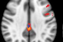
The marker most commonly used is PET imaging of brain metabolism, using FDG as a tracer. This technique shows typical hypometabolic patterns in different dementia syndromes, with an overall superior sensitivity compared with hypoperfusion assessment by SPECT. A second molecular marker used for the differential diagnosis of dementias is dopamine transporter imaging, mainly by SPECT and 123I-ioflupane, the authors explained.
Over the past decade, specific nuclear medicine tracers able to visualize in vivo amyloid plaques, which represent an important pathological hallmark of Alzheimer's disease, have been tested and validated in humans, and most amyloid imaging investigations have used C-11-Pittsburgh compound B as a tracer, they noted. This tracer has proved to be able to bind fibrillary amyloid with a good correlation with postmortem measures.
Newer F-18-labeled compounds are also becoming available, and are likely to have a wider diffusion in clinical practice, owing to their greater availability, the researchers wrote. This is linked to the longer half-life of the isotope; whereas C-11 has a half-life of 20 minutes and needs an onsite cyclotron for production of the tracer, F-18 has a half-life of 110 minutes and therefore the tracer can be produced off-site.
Looking to the future, Haller and his colleagues are studying the use of functional MRI to assess the vascular reserve of patients with MCI.
"Because of the neurovascular coupling, we hope that the assessment of the vascular reserve might be a surrogate marker of the neurocognitive reserve, which has a substantial interindividual variation and complicates the correlation between morphometric changes and cognitive status," he told AuntMinnieEurope.com.
The Geneva group is also investigating new amyloid-specific tracers for PET and assessment of per-MCI in MRI in a longitudinal design to predict conversion from control to MCI. Overall, the team's main focus is on the cerebral perfusion reserve in dementia, notably Alzheimer's disease. They think computer-aided advanced image analysis is useful for individual classification of many diseases, such as Alzheimer's and psychosis, which have associated morphologic brain changes.
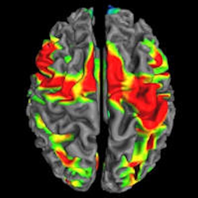
A recent study individually classified patients at risk for psychosis based on cortical thickness. Ongoing research aims to include other types of brain imaging parameters, such as white matter diffusion tensor imaging and different classification algorithms.