Lung-RADS v2022 classification tool can enable earlier diagnosis of lung cancers associated with cystic airspaces in 75% of cases, according to a study of 1,258 participants from three landmark screening trials published in <i>Radiology</i>.
- Lung cancers associated with cystic airspaces account for approximately 22.7% of missed or delayed diagnoses in low-dose CT screening
- Lung-RADS v2022 criteria could have enabled earlier diagnosis in 75% of cases (55 of 73 cystic airspace cancers) in the retrospective study
- Five-year lung cancer-specific survival was 51.4% for cystic airspace cancers compared with 53.2% for noncystic non-small cell lung cancers
- The study analyzed data from three major clinical trials involving 1,258 participants with histologically confirmed lung cancer
- Radiologist education and integration of cyst-specific assessment into nodule management guidelines is essential for reducing missed cancers of this phenotype
The Lung-RADS v2022 classification tool can facilitate earlier diagnosis of challenging lung cancers associated with cystic airspaces, according to a study published November 10 in Radiology.
A team of researchers in Italy and Germany used Lung-RADS v2022 in a secondary analysis of three landmark lung cancer screening trials involving 1,258 participants, with findings suggesting the tool has the potential to reduce delayed diagnoses.
"Application of the [low-dose] CT Lung Imaging Reporting and Data System 2022 criteria and management recommendations may facilitate earlier recognition and diagnosis of lung cancers associated with [cystic airspaces]," noted lead author Mario Mascalchi, MD, PhD, of the University of Florence, and colleagues.
Lung cancer associated with cystic airspaces is a radiologic-morphologic phenotype in which a primary lung cancer arises within, abuts, or forms the wall of a discrete air-filled space, the authors explained. The phenotype accounts for approximately 22.7% of missed or delayed lung cancer diagnoses in low-dose CT screening. Lung-RADS v2022 introduced the atypical pulmonary cyst category to address the gap, but whether its criteria would translate to earlier diagnosis had not been tested, they noted.
To bridge the gap, the group analyzed data from three registered clinical trials: the U.S. National Lung Screening Trial (NLST, 2002–2007), the Italian Lung Cancer Screening Trial (ITALUNG, 2004–2010), and the German Lung Cancer Screening Intervention trial (LUSI, 2007–2016). They reviewed all available low-dose CT examinations from 1,258 participants with histologically confirmed lung cancer.
Two radiologists jointly identified and characterized cystic airspace-associated cancers, applied Lung-RADS v2022 atypical pulmonary cyst criteria retrospectively, and analyzed lung cancer-specific survival.
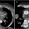
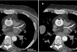
 Examples of the potential application of the Lung Imaging Reporting and Data System (Lung-RADS) 2022 criteria for earlier identification of lung cancer associated with cystic airspaces (CA) manifesting as atypical pulmonary cysts and corresponding management recommendations to anticipate diagnostic work-up for three participants from the National Lung Screening Trial. Application of Lung-RADS 2022 to serial low-dose CT (LDCT) examinations could have led to potential diagnostic anticipation in 80% (24 of 30) of unilocular cysts with associated nodule, 53% (10 of 19) of thick-walled unilocular cysts, and 88% (21 of 24) of multilocular cyst. (A) Participant 54, a 64-year-old female. Baseline LDCT scan (T0) shows a multilocular atypical pulmonary cyst (thick arrow) in the right upper lobe, consistent with a Lung-RADS 2022 category 4A (suspicious) lesion, according to which 3-month LDCT would have been recommended. At the first annual repeat examination (T1), two solid nodules (8.4 and 4.4 mm) (thin arrows) appeared within the cyst, consistent with lung cancer associated with CA and a Lung-RADS category 4B (very suspicious) lesion, according to which diagnostic CT, PET/CT, or tissue sampling would have been recommended 1 year after the baseline LDCT examination. At the next annual repeat examination (T2), the lesion reverted to a solid mass, consistent with a category 4B lesion. A screening-detected stage IV adenocarcinoma was diagnosed 2 years after the baseline LDCT examination and was the cause of death 3 months later. (B) Participant 44, a 66-year-old male. Baseline LDCT scan (T0) shows a large unilocular cyst with a small (4.8-mm), thick-walled nodule (arrow) in the right upper lobe, consistent with the features of an atypical pulmonary cyst with a thick wall and qualifying the lesion as a Lung-RADS 2022 category 4A (suspicious) lesion, according to which 3-month LDCT would have been recommended. At the first annual repeat examination (T1), the nodule had grown more than 1.5 mm, consistent with a category 4B (very suspicious) lesion according to Lung-RADS 2022; diagnostic CT, PET/CT, or tissue sampling would have been recommended 1 year after the baseline examination. At the second annual repeat examination (T2), elongated thickening of the adjacent cyst wall was observed, consistent with a category 4B lesion as per Lung-RADS 2022. A non–screening-detected stage IIA squamous cell carcinoma was ultimately diagnosed 2.6 years after the baseline LDCT examination and was the cause of death 3.3 years later. (C) Participant 43, a 70-year-old male. Baseline LDCT scan (T0) shows paraseptal emphysema areas in the right lower lobe. The first annual repeat LDCT examination (T1) shows thickening (arrow) of the airspace walls by more than 1.5 mm, consistent with an atypical pulmonary cyst and qualifying for a category 4B (very suspicious) lesion according to Lung-RADS 2022; diagnostic CT, PET/CT, or tissue sampling would have been recommended 1 year after the baseline LDCT examination. At the second annual repeat examination (T2), the solid components had almost filled the airspace. A screening-detected stage IV squamous cell carcinoma was ultimately diagnosed 2.6 years after baseline LDCT and was the cause of death 10 months later.RSNA
"Participants with lung cancers associated with cystic airspaces had 5-year cancer-specific survival similar to those with noncystic non–small cell lung cancers, and retrospective simulations suggested that management recommendations based on low-dose CT Lung Imaging Reporting and Data System 2022 enabled earlier diagnosis," the group wrote.
Prospective validation studies are now needed, and such futures studies should incorporate dedicated segmentation tools to improve the characterization of these lesions, the researchers concluded.
In an accompanying editorial, Suzanne Byrne, MD, and Mark Hammer, MD, both of Mass General Brigham and Harvard Medical School, noted that integrating cyst-specific assessment into nodule management guidelines in both screening and incidental detection settings will be essential to reducing the missed cancers of this phenotype, they added.
"The current study reinforces the need for radiologist education and integration of cyst-specific assessment into nodule management guidelines, not only in the context of lung cancer screening but also in incidental detection," Byrne and Hammer wrote.
The full study is available here.