Prof. Jacob Valk, PhD.
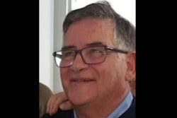
Renowned neuroradiologist Prof. Jacob Valk, PhD, died on 13 April at the age of 94 in Wilnis, a village in the Dutch province of Utrecht. He was a world expert on pediatric neuroimaging, inborn errors of metabolism, toxic encephalopathies, and white matter disorders in dementia.
For nearly 20 years, Valk was a professor of radiology and neuroradiology, and head of the Departments of Radiology and Neuroradiology at Vrije Universiteit Medical Centre, the Netherlands. After his retirement, he worked as a neuroradiologist at the MRI Center in Amsterdam.
Valk became an honorary member of the RSNA in 2003.
"Jaap Valk has excelled in the fields of neurology, psychiatry, and neuroradiology. He published a medical book every year during the middle of his career. His writings include seminal concepts on such topics as white matter disease of the brain," noted RSNA 2003 President Dr. Peggy J. Fritzsche. "He is a Renaissance man, publishing in both the arts and sciences. He is a talented pianist who has entertained many of us with his regalia of show tunes and classical pieces. He is still actively contributing, as a writer and teacher."
Valk wrote more than 220 articles and book chapters, as well as 16 books, including Magnetic Resonance of Myelin, Myelination and Myelin Disorders, a standard text in the field.
In an announcement, Edith M. de Boer described him as “erudite, creative, a gifted pianist, generous, inspirational, funny, pleasantly disturbed, an experienced world traveler, and diver.”
A farewell meeting will be held on Saturday 20 April at noon at the Crematorium Westgaarde in Amsterdam. Afterward, there will be an opportunity for attendees to meet each other in the reception room, the announcement noted. Also, the next issue of MemoRad, the Dutch radiology magazine, will feature a tribute article written by Prof. Jonas Castelijns, Prof. Frederik Barkhof, and Dr. Paul Algra.