Training in interventional radiology (IR), as well as the cost-effectiveness of IR, is coming under intense scrutiny at France's national radiology congress, JFR 2023. The Canadian approach to IR training is seen by many as a successful and viable model for Europeans to emulate.
IR training and continuing medical education (CME) is organized in close collaboration with diagnostic radiology, Dr. Gilles Soulez, an interventional radiologist at Centre Hospitalier de l'Université de Montréal (CHUM), told AuntMinnieEurope.com ahead of JFR 2023, which runs from 13 to 16 October.
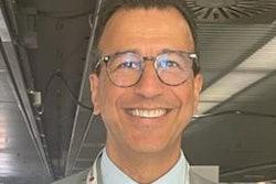
"There's always a risk of minimizing the role of diagnostic radiology in interventional radiology, which can be problematic in rural areas," noted Soulez, who will become an honorary member of the French Society of Radiology (SFR) at this week's congress and is past president of the Canadian Association of Radiologists (CAR).
In smaller hospitals, interventional radiologists also need to do diagnostic radiology. In the U.S., accessibility to IR in small practices is now problematic, he added.
How the Canadians do it
The Canadian program is "quite demanding in terms of administration and follow-up," according to Soulez, who will talk on this topic at the JFR Executive Leadership Session on 14 October. He was born in France and completed his medical studies at the Université Paris Descartes before moving to Montreal in 1991. He received the award of excellence and innovation from the Cardiovascular and Interventional Radiology Society of Europe (CIRSE) in 2013.
Soulez, who is now a professor of radiology at Université de Montréal, prepared his JFR presentation with Dr. Marie France Giroux, program director of IR residency and fellowship programs.
IR is a recognized subspeciality program, and candidates must enter via the five-year diagnostic radiology (DR) program, they explained. One year of IR training can be credited during the final three years of the DR program, and one additional year is spent specifically in IR. All candidates have a dual certification (DR and IR). IR fellowships are still provided outside the IR residency program.
"Fellows outside the IR program can pass the written and oral examination of the IR residency program," Soulez and Giroux stated. "There is an appropriate organizational structure, with leadership and administrative personnel to support the residency program, teachers, and residents effectively."
A program director with sufficient administrative support and protected time manages the program, and significant stakeholders are involved. All aspects of the residency program are collaboratively overseen by the program director and committee.
"Residents are prepared for independent practice," they continued. "The residency program has the clinical, physical, technological, and financial resources to provide all residents with the educational experiences needed to acquire all competencies and/or objectives."
The residency program provides educational experiences designed to facilitate residents' attainment of the outcomes-based competencies (competence by design curriculum) and/or objectives. Safety and wellness are promoted, and residents are treated fairly and supported adequately. There is continuous improvement of the educational experiences, to improve the residency program and ensure residents are prepared for independent practice, they pointed out.
Debate on "valorization"
At the same JFR session, attention will also focus on IR and valorization (the act or process of giving, assigning, or enhancing value). ECR 2024 President Prof. Catalano from Rome will elaborate on the European experience. Prof. Ashin Gangi, PhD, chair of radiology and nuclear medicine at Strasbourg University Hospital, France, and past president of CIRSE, will speak on the French experience.
There are not enough interventional radiologists in France, according to Gangi. "In my team, each of us is working one day per week at two other hospitals in Colmar and Mulhouse," he said in an interview at JFR 2022. "That's not enough for the 2 million population we have to cover. We are short of more than 100 to 200 interventional radiologists in France."
IR is still a relatively male-dominated subspecialty and action must be taken to improve the situation, he conceded. "65% of medical students are women," Gangi noted. "For your future, you need to do something in order to survive."
Burnout is an issue in radiology in general, but thankfully not in his group, he emphasized. "My team is made up of crazy drivers, that means we're always racing! We're lucky, we have plenty of novelty. No one is affected by burnout, but I recognize the issue at the hospital. Burnout is killing some people's motivation. COVID has made it even more obvious."
Enthusiasm is his preventive cure. "When patients see you after the treatment and they're happy, this recharges your battery," Gangi said.