PARIS - How do the public and private sectors work together in French radiology? Prof. Afshin Gangi from Strasbourg shed light on France's unique system. In a video interview at the French national radiology congress, JFR, he also speaks about how his team avoids burnout.
"France is very different from other countries. The public hospitals are 100% under the control of the French government," explained Gangi, who is head of radiology at Strasbourg University Hospital and a professor at King's College London. Radiologists, however, can work in both the public and private sectors if they allocate at least 20% of their time to the other sector, he added.
Although Gangi doesn't feel any tension exists between public and private radiologists, he acknowledges that a certain "Parisianisme" -- a tendency to separate between Paris and the rest of France -- does linger in radiology and healthcare in general. "Parisianisme is inevitable in France," he said. "It's a pretty centralized country. But even in our national university system, there are plenty of people coming from outside of the capital."
There are not enough interventional radiologists in France, according to Gangi, who is the immediate past president of the Cardiovascular and Interventional Radiological Society of Europe (CIRSE). "In my team, each of us is working one day per week at two other hospitals in Colmar and Mulhouse," he said. "That's not enough for the 2 million population we have to cover. We are short of more than 100 to 200 interventional radiologists in France."
Interventional radiology is still a relatively male-dominated subspecialty and action must be taken to improve the situation, he conceded. "65% of medical students are women," Gangi noted. "For your future, you need to do something in order to survive."
Burnout is an issue in radiology in general, but thankfully not in his group, according to Gangi. "My team is made up of crazy drivers, that means we're always racing! We're lucky, we have plenty of novelty," he said. "No one is affected by burnout, but I recognize the issue at the hospital. Burnout is killing some people's motivation. COVID has made it even more obvious."
Enthusiasm is his preventive cure. "When patients see you after the treatment and they're happy, this recharges your battery," Gangi concluded.
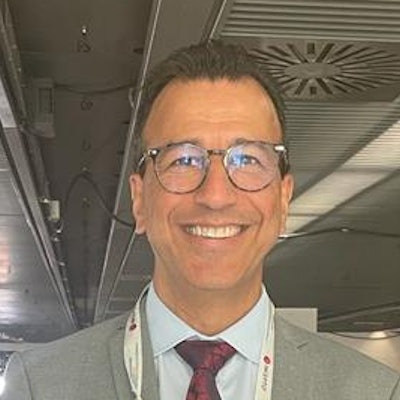
Prof. Afshin Gangi talked about the structure of radiology in France -- as well as the phenomenon of "Parisianisme" -- at the French national radiology congress, JFR.


![Examples of ultrasound findings and techniques. (A) Images in a 39-year-old male patient with a mass in the left thigh. The mass is heterogeneous on the B-mode US image (compared with the patient in D) and showed increased microvascularity (superb microvascular imaging [SMI]) and shear-wave elastography (SWE) values. Undifferentiated pleomorphic sarcoma was diagnosed at biopsy (with pleomorphic rhabdomyosarcoma in surgical specimen). (B) Images in an 18-year-old male patient with a mass in the left leg. The mass is hypoechoic on the B-mode image, with no other findings suggestive of malignancy. The lesion is in contact with the cortex of the tibia, which is slightly irregular. CT revealed a doubtful anteromedial tibial erosion. The microvascular study demonstrated high vascularization, suggestive of malignancy. Periosteal Ewing sarcoma was diagnosed with both histologic and immunohistochemical confirmation. (C) Images in a 69-year-old female patient with a lump growing on the outside of the left leg. Multiple SWE examinations were performed (please note the high values obtained in the measurements, whereas the color map highlights the stiffness relative to adjacent tissues). SMI showed areas of increased vascularization to target for sampling. Undifferentiated spindle cell sarcoma was diagnosed at biopsy, with residual leiomyosarcoma in the surgical specimen after neoadjuvant therapy. (D) Images in a 56-year-old female patient with a mass in the right thigh. The mass is heterogeneous at both B-mode ultrasound (similar to patient A) and MRI (coronal T2-weighted spectral attenuated inversion recovery [SPAIR]; T1-weighted pre-contrast and postcontrast imaging), which even shows uptake after the administration of paramagnetic contrast material, which is traditionally suggestive of malignancy. Low values at SMI and elastography are suggestive of benignity. Spindle cell lipoma was diagnosed at biopsy, with atypical spindle cell lipomatous tumor in the surgical specimen.](https://img.auntminnieeurope.com/mindful/smg/workspaces/default/uploads/2026/08/images-radiol250278fig2.APCFLSvX6p.jpg?auto=format%2Ccompress&fit=crop&h=100&q=70&w=100)


![Examples of ultrasound findings and techniques. (A) Images in a 39-year-old male patient with a mass in the left thigh. The mass is heterogeneous on the B-mode US image (compared with the patient in D) and showed increased microvascularity (superb microvascular imaging [SMI]) and shear-wave elastography (SWE) values. Undifferentiated pleomorphic sarcoma was diagnosed at biopsy (with pleomorphic rhabdomyosarcoma in surgical specimen). (B) Images in an 18-year-old male patient with a mass in the left leg. The mass is hypoechoic on the B-mode image, with no other findings suggestive of malignancy. The lesion is in contact with the cortex of the tibia, which is slightly irregular. CT revealed a doubtful anteromedial tibial erosion. The microvascular study demonstrated high vascularization, suggestive of malignancy. Periosteal Ewing sarcoma was diagnosed with both histologic and immunohistochemical confirmation. (C) Images in a 69-year-old female patient with a lump growing on the outside of the left leg. Multiple SWE examinations were performed (please note the high values obtained in the measurements, whereas the color map highlights the stiffness relative to adjacent tissues). SMI showed areas of increased vascularization to target for sampling. Undifferentiated spindle cell sarcoma was diagnosed at biopsy, with residual leiomyosarcoma in the surgical specimen after neoadjuvant therapy. (D) Images in a 56-year-old female patient with a mass in the right thigh. The mass is heterogeneous at both B-mode ultrasound (similar to patient A) and MRI (coronal T2-weighted spectral attenuated inversion recovery [SPAIR]; T1-weighted pre-contrast and postcontrast imaging), which even shows uptake after the administration of paramagnetic contrast material, which is traditionally suggestive of malignancy. Low values at SMI and elastography are suggestive of benignity. Spindle cell lipoma was diagnosed at biopsy, with atypical spindle cell lipomatous tumor in the surgical specimen.](https://img.auntminnieeurope.com/mindful/smg/workspaces/default/uploads/2026/08/images-radiol250278fig2.APCFLSvX6p.jpg?auto=format%2Ccompress&fit=crop&h=112&q=70&w=112)