
The European Society of Radiology (ESR) has revealed plans to take its interactive learning Escape Rooms project on tour this autumn at two national congresses.
The Escape Rooms were among the most popular interactive highlights at ECR 2022 and 2023, being run by radiologists from the Charité in Berlin, ESR announced on 6 July. Teams taking part in challenges act as first-year radiology residents on their first night shift when the trauma team brings in a young patient in a car accident. Participants must complete the diagnoses within a given time frame so that the surgical team knows what to do in the operating room.
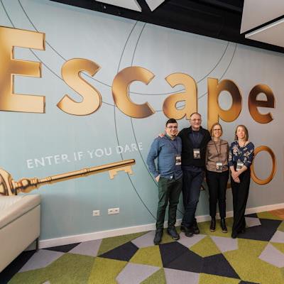
The ESR Escape Room initiative made a significant impact at ECR 2023. Photo courtesy of Sebastian Kreuzberger/ESR.The Escape Rooms will be hosted at the Journées Francophones de Radiologie (JFR) 2023 and the Romanian Congress of Radiology and Imaging (SRIM) 2023, the ESR said. The Berlin team will again be in charge of the project.
In other news, the ESR reported new impact factors for its journals. European Radiology Experimental has received its first-ever impact factor of 3.8. In addition, European Radiology, the organization's flagship publication for clinical science, received a 2022 impact factor of 5.9 and Insights into Imaging, dedicated to critical and educational articles, received a 2022 impact factor of 4.7


![Overview of the study design. (A) The fully automated deep learning framework was developed to estimate body composition (BC) (defined as subcutaneous adipose tissue [SAT] in liters; visceral adipose tissue [VAT] in liters; skeletal muscle [SM] in liters; SM fat fraction [SMFF] as a percentage; and intramuscular adipose tissue [IMAT] in deciliters) from MRI. The fully automated framework comprised one model (model 1) to quantify different BC measures (SAT, VAT, SM, SMFF, and IMAT) as three-dimensional (3D) measures from whole-body MRI scans. The second model (model 2) was trained to identify standardized anatomic landmarks along the craniocaudal body axis (z coordinate field), which allowed for subdividing the whole-body measures into different subregions typically examined on clinical routine MRI scans (chest, abdomen, and pelvis). (B) BC was quantified from whole-body MRI in over 66,000 individuals from two large population-based cohort studies, the UK Biobank (UKB) (36,317 individuals) and the German National Cohort (NAKO) (30,291 individuals). Bar graphs show age distribution by sex and cohort. BMI = body mass index. (C) After the performance assessment of the fully automated framework, the change in BC measures, distributions, and profiles across age decades were investigated. Age-, sex-, and height-adjusted body composition reference curves were calculated and made publicly available in a web-based z-score calculator (https://circ-ml.github.io).](https://img.auntminnieeurope.com/mindful/smg/workspaces/default/uploads/2026/05/body-comp.XgAjTfPj1W.jpg?auto=format%2Ccompress&fit=crop&h=112&q=70&w=112)


