The case of a two-year-old girl with a button battery lodged in her esophagus for 12 months highlights the urgent need for imaging examinations in patients with a history of difficulty in swallowing, doctors from Tanzania have reported.
A team of surgeons at Kilimanjaro Christian Medical Centre in Moshi removed a button battery from the child by thoracotomy after identifying the object on x-ray and CT exams.
"Luckily the battery case was intact during removal, preventing overt toxic alkaline hydrolysis from its contacts," wrote corresponding author Dr. Kennedy Misso and colleagues, in an article published on 20 August in the International Journal of Surgery Case Reports.
Foreign body ingestion is frequent among children under four years old, with button batteries among the most common objects involved, the authors wrote.
In this case, a two-year-old girl presented with a one-year history of progressive difficulty in swallowing. She had experienced unintentional weight loss of nearly two kilograms in recent months and had been recently treated empirically for pneumonia at nearby facilities without significant relief, the authors noted.
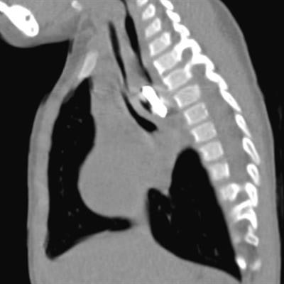
Posteroanterior chest x-ray and sagittal view of the thoracic CT revealing a foreign body in the thoracic esophagus. Image courtesy of the International Journal of Surgery Case Reports through CC BY 4.0.A chest x-ray revealed a halo sign and a thoracic CT scan showed a round object obstructing her esophagus. Attempts to visualize and retrieve the foreign body endoscopically were unsuccessful, and the surgeons performed a thoracotomy to retrieve it.
"A month later, she had gained two kilograms, she was playful, and feeding normally," the group wrote.
The authors noted that the battery was removed within a "false diverticulum" – a pouch in the child's esophagus that had formed due to prolonged esophageal irritation and intraluminal pressure. Typically, in pediatrics, diverticula are rare and typically stem from congenital anomalies.
"A high suspicion index and radiological investigations are needed when evaluating children with respiratory tract infections. Early diagnosis is the key to achieving successful endoscopic removal with minimal morbidity," the group concluded.
The full case report can be found here.

![Examples of ultrasound findings and techniques. (A) Images in a 39-year-old male patient with a mass in the left thigh. The mass is heterogeneous on the B-mode US image (compared with the patient in D) and showed increased microvascularity (superb microvascular imaging [SMI]) and shear-wave elastography (SWE) values. Undifferentiated pleomorphic sarcoma was diagnosed at biopsy (with pleomorphic rhabdomyosarcoma in surgical specimen). (B) Images in an 18-year-old male patient with a mass in the left leg. The mass is hypoechoic on the B-mode image, with no other findings suggestive of malignancy. The lesion is in contact with the cortex of the tibia, which is slightly irregular. CT revealed a doubtful anteromedial tibial erosion. The microvascular study demonstrated high vascularization, suggestive of malignancy. Periosteal Ewing sarcoma was diagnosed with both histologic and immunohistochemical confirmation. (C) Images in a 69-year-old female patient with a lump growing on the outside of the left leg. Multiple SWE examinations were performed (please note the high values obtained in the measurements, whereas the color map highlights the stiffness relative to adjacent tissues). SMI showed areas of increased vascularization to target for sampling. Undifferentiated spindle cell sarcoma was diagnosed at biopsy, with residual leiomyosarcoma in the surgical specimen after neoadjuvant therapy. (D) Images in a 56-year-old female patient with a mass in the right thigh. The mass is heterogeneous at both B-mode ultrasound (similar to patient A) and MRI (coronal T2-weighted spectral attenuated inversion recovery [SPAIR]; T1-weighted pre-contrast and postcontrast imaging), which even shows uptake after the administration of paramagnetic contrast material, which is traditionally suggestive of malignancy. Low values at SMI and elastography are suggestive of benignity. Spindle cell lipoma was diagnosed at biopsy, with atypical spindle cell lipomatous tumor in the surgical specimen.](https://img.auntminnieeurope.com/mindful/smg/workspaces/default/uploads/2026/08/images-radiol250278fig2.APCFLSvX6p.jpg?auto=format%2Ccompress&fit=crop&h=100&q=70&w=100)


![Examples of ultrasound findings and techniques. (A) Images in a 39-year-old male patient with a mass in the left thigh. The mass is heterogeneous on the B-mode US image (compared with the patient in D) and showed increased microvascularity (superb microvascular imaging [SMI]) and shear-wave elastography (SWE) values. Undifferentiated pleomorphic sarcoma was diagnosed at biopsy (with pleomorphic rhabdomyosarcoma in surgical specimen). (B) Images in an 18-year-old male patient with a mass in the left leg. The mass is hypoechoic on the B-mode image, with no other findings suggestive of malignancy. The lesion is in contact with the cortex of the tibia, which is slightly irregular. CT revealed a doubtful anteromedial tibial erosion. The microvascular study demonstrated high vascularization, suggestive of malignancy. Periosteal Ewing sarcoma was diagnosed with both histologic and immunohistochemical confirmation. (C) Images in a 69-year-old female patient with a lump growing on the outside of the left leg. Multiple SWE examinations were performed (please note the high values obtained in the measurements, whereas the color map highlights the stiffness relative to adjacent tissues). SMI showed areas of increased vascularization to target for sampling. Undifferentiated spindle cell sarcoma was diagnosed at biopsy, with residual leiomyosarcoma in the surgical specimen after neoadjuvant therapy. (D) Images in a 56-year-old female patient with a mass in the right thigh. The mass is heterogeneous at both B-mode ultrasound (similar to patient A) and MRI (coronal T2-weighted spectral attenuated inversion recovery [SPAIR]; T1-weighted pre-contrast and postcontrast imaging), which even shows uptake after the administration of paramagnetic contrast material, which is traditionally suggestive of malignancy. Low values at SMI and elastography are suggestive of benignity. Spindle cell lipoma was diagnosed at biopsy, with atypical spindle cell lipomatous tumor in the surgical specimen.](https://img.auntminnieeurope.com/mindful/smg/workspaces/default/uploads/2026/08/images-radiol250278fig2.APCFLSvX6p.jpg?auto=format%2Ccompress&fit=crop&h=112&q=70&w=112)