Photon-counting CT (PCCT) effectively detects heart disease in high-risk patients, offering "significant benefit for people previously ineligible for noninvasive screening," researchers from the have reported in a study published on 20 June in Radiology.
The findings could help clinicians better diagnose heart disease and tailor patient care, lead author Dr. Muhammad Hagar, from the University of Freiburg in Germany, told AuntMinnieEurope.com.
"The results broaden the spectrum of patients who can benefit from noninvasive coronary CT angiography, offering a safer and less stressful experience for them," he said. "Moreover, if confirmed in other trials, it may change the current guidelines that don't recommend [coronary CT angiography] for high-risk individuals."
Coronary artery disease is the most common form of heart disease, according to Hagar and colleagues. Coronary CT angiography (CCTA) is effective for ruling the disease out in patients at low- or intermediate risk, but using it in a high-risk population is difficult because many of these patients have coronary calcifications or stents; the calcifications often look bigger than they actually are, translating to the overestimation of blocks and plaque in the heart vessels and false-positive results, according to Hagar.
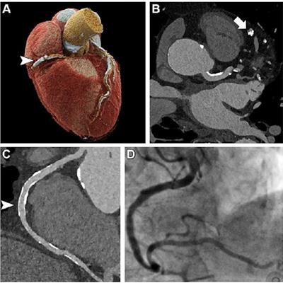
 Ultrahigh-resolution (UHR) coronary CT angiography (CCTA) in an 85-year-old man before transcatheter aortic valve replacement. Despite a stent in the right coronary artery and very severe coronary sclerosis with an Agatston score of 4,162, diagnostic visualization of the coronary arteries succeeded, and obstructive coronary artery disease was excluded on CT images. (A) Three-dimensional cinematic rendering of the heart. The stent (arrowhead) is visible in the middle segment of the right coronary artery. (B) UHR CCTA with 0.2-mm axial sections. The lumen (arrow) of the severely calcified distal left anterior descending artery can be assessed without artifacts. (C) Curved multiplanar reformations of the right coronary artery with a diagnostic display of the stent lumen (arrowhead). (D) Invasive coronary angiography enables exclusion of in-stent stenosis. Image and caption courtesy of the RSNA.
But the advent of ultrahigh-resolution coronary CT angiography conducted with a PCCT scanner shows promise for changing the game, the investigators wrote. To confirm this, they conducted a study that included 68 patients and compared the diagnostic accuracy of ultra-high-resolution CCTA with the standard of care, which is invasive coronary angiography. Study participants had severe aortic valve stenosis.
The group found that ultrahigh-resolution CCTA was highly sensitive and specific for identifying coronary artery disease. It garnered an overall image quality score of 1.5 on a five-point scale (with 1 being excellent and 5 nondiagnostic).
| Performance of ultrahigh resolution CCTA with PCCT for detecting heart disease | ||
| Measure | Stenosis of ≥ 50% | Stenosis of ≥ 70% |
| Sensitivity | 96% | 100% |
| Specificity | 84% | 76% |
| Positive predictive value | 77% | 60% |
| Negative predictive value | 97% | 60% |
| Accuracy | 88% | 82% |
PCCT shows promise for improving how patients at high-risk of heart disease are diagnosed, according to Hagar. He and his fellow researchers expect PCCT technology to enter mainstream clinical practice over the next decade, and plan to further investigate its diagnostic capability for other applications, such as cancer imaging.
"It appears that the spectrum of patients benefiting from undergoing noninvasive CCTA has been significantly broadened by photon-counting detector technology," Hagar said. "This is excellent news for these patients and the imaging community."
More research is needed, wrote Michelle Williams, MDchB, PhD, and David Newby, DM, PhD, both of the University of Edinburgh in Scotland, in an accompanying editorial.
"The use of PCCT scanners offers several potential advances for CT imaging," they wrote. "However, at present, we do not know which patients would benefit most from this technology or how best to use it in clinical practice."

![Examples of ultrasound findings and techniques. (A) Images in a 39-year-old male patient with a mass in the left thigh. The mass is heterogeneous on the B-mode US image (compared with the patient in D) and showed increased microvascularity (superb microvascular imaging [SMI]) and shear-wave elastography (SWE) values. Undifferentiated pleomorphic sarcoma was diagnosed at biopsy (with pleomorphic rhabdomyosarcoma in surgical specimen). (B) Images in an 18-year-old male patient with a mass in the left leg. The mass is hypoechoic on the B-mode image, with no other findings suggestive of malignancy. The lesion is in contact with the cortex of the tibia, which is slightly irregular. CT revealed a doubtful anteromedial tibial erosion. The microvascular study demonstrated high vascularization, suggestive of malignancy. Periosteal Ewing sarcoma was diagnosed with both histologic and immunohistochemical confirmation. (C) Images in a 69-year-old female patient with a lump growing on the outside of the left leg. Multiple SWE examinations were performed (please note the high values obtained in the measurements, whereas the color map highlights the stiffness relative to adjacent tissues). SMI showed areas of increased vascularization to target for sampling. Undifferentiated spindle cell sarcoma was diagnosed at biopsy, with residual leiomyosarcoma in the surgical specimen after neoadjuvant therapy. (D) Images in a 56-year-old female patient with a mass in the right thigh. The mass is heterogeneous at both B-mode ultrasound (similar to patient A) and MRI (coronal T2-weighted spectral attenuated inversion recovery [SPAIR]; T1-weighted pre-contrast and postcontrast imaging), which even shows uptake after the administration of paramagnetic contrast material, which is traditionally suggestive of malignancy. Low values at SMI and elastography are suggestive of benignity. Spindle cell lipoma was diagnosed at biopsy, with atypical spindle cell lipomatous tumor in the surgical specimen.](https://img.auntminnieeurope.com/mindful/smg/workspaces/default/uploads/2026/08/images-radiol250278fig2.APCFLSvX6p.jpg?auto=format%2Ccompress&fit=crop&h=100&q=70&w=100)






![Examples of ultrasound findings and techniques. (A) Images in a 39-year-old male patient with a mass in the left thigh. The mass is heterogeneous on the B-mode US image (compared with the patient in D) and showed increased microvascularity (superb microvascular imaging [SMI]) and shear-wave elastography (SWE) values. Undifferentiated pleomorphic sarcoma was diagnosed at biopsy (with pleomorphic rhabdomyosarcoma in surgical specimen). (B) Images in an 18-year-old male patient with a mass in the left leg. The mass is hypoechoic on the B-mode image, with no other findings suggestive of malignancy. The lesion is in contact with the cortex of the tibia, which is slightly irregular. CT revealed a doubtful anteromedial tibial erosion. The microvascular study demonstrated high vascularization, suggestive of malignancy. Periosteal Ewing sarcoma was diagnosed with both histologic and immunohistochemical confirmation. (C) Images in a 69-year-old female patient with a lump growing on the outside of the left leg. Multiple SWE examinations were performed (please note the high values obtained in the measurements, whereas the color map highlights the stiffness relative to adjacent tissues). SMI showed areas of increased vascularization to target for sampling. Undifferentiated spindle cell sarcoma was diagnosed at biopsy, with residual leiomyosarcoma in the surgical specimen after neoadjuvant therapy. (D) Images in a 56-year-old female patient with a mass in the right thigh. The mass is heterogeneous at both B-mode ultrasound (similar to patient A) and MRI (coronal T2-weighted spectral attenuated inversion recovery [SPAIR]; T1-weighted pre-contrast and postcontrast imaging), which even shows uptake after the administration of paramagnetic contrast material, which is traditionally suggestive of malignancy. Low values at SMI and elastography are suggestive of benignity. Spindle cell lipoma was diagnosed at biopsy, with atypical spindle cell lipomatous tumor in the surgical specimen.](https://img.auntminnieeurope.com/mindful/smg/workspaces/default/uploads/2026/08/images-radiol250278fig2.APCFLSvX6p.jpg?auto=format%2Ccompress&dpr=2&fit=crop&h=167&q=70&w=250)


