F-18 sodium fluoride (NaF) PET/CT may be a valuable tool for imaging patients with psoriatic arthritis (PsA), as it reveals new bone formation in peripheral joints missed on clinical evaluation, according to a Dutch group.
"F-18 NaF PET/CT may be a novel clinically valuable tool to detect whole-body disease activity of PsA reflected by new bone formation in all disease domains of PsA, depicted in one scan," a team led by doctoral candidate Jerney de Jongh wrote. The study was published 12 November in the European Journal of Nuclear Medicine and Molecular Imaging.
PsA is a chronic and inflammatory musculoskeletal disease associated with psoriasis and several musculoskeletal manifestations, especially enthesitis, which is a painful inflammation at the site where tendons or ligaments attach to bone. Enthesitis activity in PsA can be accompanied by new bone formation.
Clinical assessment of enthesitis is challenging and has limited accuracy, as it is based only on the presence of tenderness and general soft tissue swelling, according to the authors. Moreover, other imaging approaches such as ultrasound and MRI have proven limited in identifying these sites, they wrote.
In this study, the researchers enrolled 16 patients with a clinical diagnosis of psoriatic arthritis from a rheumatology center at Amsterdam UMC. Patients underwent whole-body PET/CT scans 45 minutes after injection with F-18 NaF, a radiotracer that can identify new bone formation based on osteoblastic activity.
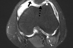
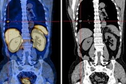
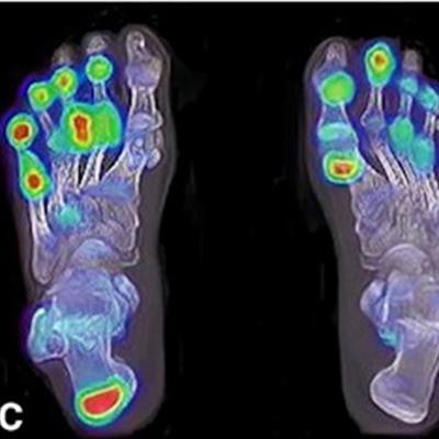
F-18 NaF enhancement in the right wrist and proximal interphalangeal joints of the hands (A), at the patella tendon insertion (B), in the metatarsophalangeal and interphalangeal joints of the feet and the right Achilles tendon (C), and in the distal interphalangeal joints of the hands (D). Image courtesy of the European Journal of Nuclear Medicine and Molecular Imaging.According to the study results, 81.8% of PET-positive joints were missed on clinical assessment. The team found that only 18.2% of PET-positive joints were clinically positive (that is, tender or swollen). But most PET-negative sites were also clinically negative (611/638). Similar results were found for entheses, with 70.5% of the PET-positive entheses missed on clinical evaluation and 29.5% of PET-positive entheses clinically identified.
De Jongh and colleagues acknowledged that their study cohort was small, and they urged further research.
"The clinical relevance of asymptomatic PET lesions in PsA should be further addressed in longitudinal studies, relating PET outcome with clinical and radiological follow-up over time," the investigators concluded.


![Examples of ultrasound findings and techniques. (A) Images in a 39-year-old male patient with a mass in the left thigh. The mass is heterogeneous on the B-mode US image (compared with the patient in D) and showed increased microvascularity (superb microvascular imaging [SMI]) and shear-wave elastography (SWE) values. Undifferentiated pleomorphic sarcoma was diagnosed at biopsy (with pleomorphic rhabdomyosarcoma in surgical specimen). (B) Images in an 18-year-old male patient with a mass in the left leg. The mass is hypoechoic on the B-mode image, with no other findings suggestive of malignancy. The lesion is in contact with the cortex of the tibia, which is slightly irregular. CT revealed a doubtful anteromedial tibial erosion. The microvascular study demonstrated high vascularization, suggestive of malignancy. Periosteal Ewing sarcoma was diagnosed with both histologic and immunohistochemical confirmation. (C) Images in a 69-year-old female patient with a lump growing on the outside of the left leg. Multiple SWE examinations were performed (please note the high values obtained in the measurements, whereas the color map highlights the stiffness relative to adjacent tissues). SMI showed areas of increased vascularization to target for sampling. Undifferentiated spindle cell sarcoma was diagnosed at biopsy, with residual leiomyosarcoma in the surgical specimen after neoadjuvant therapy. (D) Images in a 56-year-old female patient with a mass in the right thigh. The mass is heterogeneous at both B-mode ultrasound (similar to patient A) and MRI (coronal T2-weighted spectral attenuated inversion recovery [SPAIR]; T1-weighted pre-contrast and postcontrast imaging), which even shows uptake after the administration of paramagnetic contrast material, which is traditionally suggestive of malignancy. Low values at SMI and elastography are suggestive of benignity. Spindle cell lipoma was diagnosed at biopsy, with atypical spindle cell lipomatous tumor in the surgical specimen.](https://img.auntminnieeurope.com/mindful/smg/workspaces/default/uploads/2026/08/images-radiol250278fig2.APCFLSvX6p.jpg?auto=format%2Ccompress&fit=crop&h=100&q=70&w=100)


![Examples of ultrasound findings and techniques. (A) Images in a 39-year-old male patient with a mass in the left thigh. The mass is heterogeneous on the B-mode US image (compared with the patient in D) and showed increased microvascularity (superb microvascular imaging [SMI]) and shear-wave elastography (SWE) values. Undifferentiated pleomorphic sarcoma was diagnosed at biopsy (with pleomorphic rhabdomyosarcoma in surgical specimen). (B) Images in an 18-year-old male patient with a mass in the left leg. The mass is hypoechoic on the B-mode image, with no other findings suggestive of malignancy. The lesion is in contact with the cortex of the tibia, which is slightly irregular. CT revealed a doubtful anteromedial tibial erosion. The microvascular study demonstrated high vascularization, suggestive of malignancy. Periosteal Ewing sarcoma was diagnosed with both histologic and immunohistochemical confirmation. (C) Images in a 69-year-old female patient with a lump growing on the outside of the left leg. Multiple SWE examinations were performed (please note the high values obtained in the measurements, whereas the color map highlights the stiffness relative to adjacent tissues). SMI showed areas of increased vascularization to target for sampling. Undifferentiated spindle cell sarcoma was diagnosed at biopsy, with residual leiomyosarcoma in the surgical specimen after neoadjuvant therapy. (D) Images in a 56-year-old female patient with a mass in the right thigh. The mass is heterogeneous at both B-mode ultrasound (similar to patient A) and MRI (coronal T2-weighted spectral attenuated inversion recovery [SPAIR]; T1-weighted pre-contrast and postcontrast imaging), which even shows uptake after the administration of paramagnetic contrast material, which is traditionally suggestive of malignancy. Low values at SMI and elastography are suggestive of benignity. Spindle cell lipoma was diagnosed at biopsy, with atypical spindle cell lipomatous tumor in the surgical specimen.](https://img.auntminnieeurope.com/mindful/smg/workspaces/default/uploads/2026/08/images-radiol250278fig2.APCFLSvX6p.jpg?auto=format%2Ccompress&fit=crop&h=112&q=70&w=112)