A novel MRI registration method can track changes in intervertebral discs noninvasively while characterizing disc structures, according to research presented on 10 May at the annual meeting of the International Society for Magnetic Resonance in Medicine (ISMRM).
In her presentation, Dr. Frida Johansson from the University of Gothenburg in Sweden described her team's research, which found that deformation of the intervertebral disc (IVD) in the spine depends on disc degeneration and lumbar spine level. The team used a technique called deformation-field MRI for their findings.
"Deformation-field MRI seems to be a promising tool to study the degeneration process of the disc and might be useful in evaluating painful disc conditions," Johansson said.
ISMRM is holding this week's meeting in conjunction with the European Society for Magnetic Resonance in Medicine and Biology and the International Society for MR Radiographers and Technologists.
Spreading the load
The intervertebral disc distributes the compressive load on the spine, acting as a shock absorber. Disc tissue degenerates during the aging process, which causes the disc to lose its ability to withhold pressure. This, in turn, leads to increased back pain.
It remains unknown how the IVD responds to mechanical stimuli in vivo. Johansson said experiments outside the body can't fully replace in vivo studies since they don't take the native environmental factors of the disc into account, such as spinal column placement, muscles surrounding the disc, disc angulation, and spinal curvature. In vivo studies could give better insight into the pathophysiology of the IVD and may improve spinal diagnostics, she added.
MRI is a nonionizing and noninvasive tool that could be used for vivo evaluation. Johansson said MRI performed during axial loading of the spine could reveal important intradiscal deformation patterns.
Johansson and colleagues wanted to find out how the lumbar IVD structure deforms in vivo during spinal loading. They used a novel, noninvasive method utilizing MRI and image registration to quantify their data.
The researchers collected MR images of the lumbar spine from 24 patients suffering from chronic low back pain, as well as 12 control patients, with a 1.5-tesla MRI scanner. They also collected T2-weighted images with the spine in unloaded and loaded states to look at IVD properties at different spinal levels.
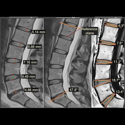
 Research presented at the annual ISMRM says that a novel MRI registration method can track changes in intervertebral discs noninvasively while characterizing disc structures. These images are used to create heat maps showing compression on different parts of the spine. Image courtesy of Dr. Frida Johansson.
Lumbar spine images acquired during axial loading were rigidly and nonrigidly transformed to corresponding unloaded T2-weighted MR images. Registration was performed through a rigid Euler transformation and a non-rigid B-spline transformation.
The team found differences in deformation for different spine levels. Johansson said that IVDs at the lower lumbar levels displayed more compression (p < 0.001), even when correcting for degeneration.
The novel imaging method displayed regional deformation variations over the IVD. The researchers found generally more compression at the middle of the IVD and a slightly left asymmetric pattern. They also found that IVDs with advanced degeneration displayed more compression at the posterior region (p = 0.035-0.045), even when correcting for spine level. These were more frequently found in the lower lumbar spine levels.
The team also noted the presence of asymmetries in the deformation pattern over the IVDs. They wrote that this was "most probably" caused by varying disc height across the IVD.
Johansson added that no significant difference was found in deformation between patients and controls.
The team concluded that while deformation-field MRI could not differentiate between symptomatic patients and asymptomatic controls on a group level, it may give individualized diagnostics when used in combination with conventional MRI markers.