
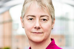
The U.K. Royal College of Radiologists (RCR) has appointed Dr. Tom Roques as vice president for clinical oncology and Dr. Priya Suresh as medical director for education and training for clinical radiology.
Tom Roques.Roques previously served as medical director for professional practice for clinical oncology at the RCR. He led clinical oncology faculty activities that guide and support oncologists throughout their careers, such as national guideline development, audit, and quality improvement. He worked as the oncology service director in Norwich from 2010 to 2016 and has been chair of the Anglia East head and neck cancer and thyroid cancer multidisciplinary teams since 2005.
Suresh first became involved at the RCR in 2011 and has served on several committees, including the Curriculum Committee, Specialty Training Board, and Faculty Board. She chaired the FRCR 2a MSK SBA and is currently the lead for the iRefer musculoskeletal guidelines.
She is also the academy lead for the Peninsula Radiology Academy, training program director and teaching lead for imaging, and honorary university fellow. Her interests in radiology include simulation training, intervention, and team building.


![Overview of the study design. (A) The fully automated deep learning framework was developed to estimate body composition (BC) (defined as subcutaneous adipose tissue [SAT] in liters; visceral adipose tissue [VAT] in liters; skeletal muscle [SM] in liters; SM fat fraction [SMFF] as a percentage; and intramuscular adipose tissue [IMAT] in deciliters) from MRI. The fully automated framework comprised one model (model 1) to quantify different BC measures (SAT, VAT, SM, SMFF, and IMAT) as three-dimensional (3D) measures from whole-body MRI scans. The second model (model 2) was trained to identify standardized anatomic landmarks along the craniocaudal body axis (z coordinate field), which allowed for subdividing the whole-body measures into different subregions typically examined on clinical routine MRI scans (chest, abdomen, and pelvis). (B) BC was quantified from whole-body MRI in over 66,000 individuals from two large population-based cohort studies, the UK Biobank (UKB) (36,317 individuals) and the German National Cohort (NAKO) (30,291 individuals). Bar graphs show age distribution by sex and cohort. BMI = body mass index. (C) After the performance assessment of the fully automated framework, the change in BC measures, distributions, and profiles across age decades were investigated. Age-, sex-, and height-adjusted body composition reference curves were calculated and made publicly available in a web-based z-score calculator (https://circ-ml.github.io).](https://img.auntminnieeurope.com/mindful/smg/workspaces/default/uploads/2026/05/body-comp.XgAjTfPj1W.jpg?auto=format%2Ccompress&fit=crop&h=112&q=70&w=112)


