A Dutch study found that adding FDG-PET metabolic imaging to contrast-enhanced CT scans significantly improved early detection of local tumor progression after colorectal liver metastasis treatment, achieving 100% sensitivity compared to 46% with CT alone, though it resulted in some false-positive findings.
- FDG-PET/contrast-enhanced CT detected all 13 cases of local progression after thermal ablation, compared to only 6 detected by CT alone (100% vs 46% sensitivity)
- After hepatic resection, the combined imaging identified all 5 cases of local progression versus 2 with CT alone (100% vs 40% sensitivity)
- Diagnostic accuracy improved substantially with combined imaging: area under the curve rose from 0.73 to 0.97 after ablation and 0.69 to 0.96 after resection
- The combined protocol identified extrahepatic disease in 19 of 22 affected patients compared to 16 with CT alone
- Trade-offs included increased false-positive findings, reducing specificity from 100% to 94% after ablation and positive predictive value from 67% to 50% after resection
A Dutch cohort study found that adding metabolic imaging to contrast-enhanced CT improved early detection of local tumor progression after liver ablation or resection, although the gain in sensitivity came with additional false-positive findings.
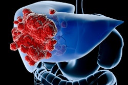
Adding F-18 FDG-PET to contrast-enhanced CT substantially improved the early detection of local tumor progression after treatment of colorectal liver metastases.
This is the core finding of a Dutch single-center cohort study published in European Radiology. Dr. Okker D. Bijlstra of Leiden University Medical Center in Leiden, the Netherlands, and Sietse van Mossel, PhD, of Leiden University Medical Center and the University of Twente in Enschede, the Netherlands, who contributed equally as co-first authors, and colleagues evaluated 64 patients with 154 treated liver lesions.
Patients underwent thermal ablation, hepatic resection, or a combination of both. They received contrast-enhanced CT and FDG-PET/contrast-enhanced CT three to five months after treatment. The researchers compared the combined examination with standard surveillance using CT and serum carcinoembryonic antigen (CEA).
A different point in care
An earlier Egyptian study covered by AuntMinnie examined whether FDG-PET/CT could distinguish responders from nonresponders during chemotherapy for metastatic colorectal cancer.
The Dutch study is a different kettle of fish. It focuses on surveillance after local treatment and asks whether metabolic imaging can expose recurrence around an ablation zone or surgical margin.
That distinction matters because fibrosis, necrosis, inflammation, and other post-treatment changes may resemble viable tumor, or conceal it, on morphological imaging. FDG-PET adds information about glucose metabolism, potentially highlighting active disease within structurally ambiguous tissue.
Timing matters
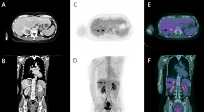
Local tumor progression occurred in 13 of 93 lesions treated with thermal ablation. Contrast-enhanced CT identified six, giving a sensitivity of 46%. FDG-PET/contrast-enhanced CT detected all 13, raising sensitivity to 100%.
Diagnostic accuracy increased from an area under the curve (AUC) of 0.73 with CT alone to 0.97 with the combined examination. Put plainly, metabolic imaging picked up progression that structural imaging left on the table.
A similar pattern emerged after hepatic resection. Five of 61 treated lesions showed local progression. CT detected two, while the combined protocol identified all five. Sensitivity increased from 40% to 100%, and the AUC rose from 0.69 to 0.96.
FDG-PET/contrast-enhanced CT also identified extrahepatic disease in 19 of 22 affected patients, compared with 16 using CT alone. However, it offered no statistically significant advantage for detecting new liver metastases away from the treated site.
The other side of the ledger
The sensitivity gains were not free of trade-offs. After ablation, the combined examination produced five false-positive findings, reducing specificity from 100% to 94% and positive predictive value from 100% to 72%. Following resection, positive predictive value fell from 67% with CT to 50% with PET/CECT.
CEA was also a poor safety net. It was false negative in 18 of 42 patients with disease progression and achieved an AUC area of 0.60. The rub is clear: low CEA cannot reliably rule out recurrence.
What the study cannot yet show
The cohort was relatively small and heterogeneous, and histological confirmation was available in only one patient. Most diagnoses were based on multidisciplinary consensus and subsequent imaging follow-up.
The authors called for prospective research to determine whether improved detection translates into better outcomes and is cost-effective. Still, FDG-PET/contrast-enhanced CT may help clinicians identify local progression earlier, while further curative treatment remains on the cards.
The study received no funding, and the authors declared no relevant company relationships.
The full study can be found here.