The British Journal of Radiology has published a collection of articles about female genitourinary oncology.
The articles explore current topics, including ovarian and cervical cancer staging, characterization, common pitfalls of the endometrium, molecular imaging, pelvic exenteration, radiogenomics in gynecological cancers, and differentiating uterine leiomyomas and leiomyosarcomas.
The special focus was guest edited by Prof. Evis Sala (University of Cambridge School of Clinical Medicine and Addenbrooke’s Hospital, Cambridge University Hospitals NHS Foundation Trust, U.K.), Dr. Stephanie Nougaret (Cancer Institute of Montpellier, France), and Dr. Alberto Vargas (Memorial Sloan Kettering Cancer Center, New York).
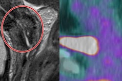
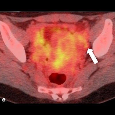
 43-year-old woman. The tumor crawls on the surface of the anterior lip of the cervix to anterior fornix of vagina. (b) Sagittal and (c) axial diffusion-weighted imaging (DWI) clearly show tumor extension to the anterior vaginal fornix. Vaginal wall invasion was suspected at anterior fornix by irregularity. This case was diagnosed as stage IIA in 2017 under the previous International Federation of Gynecology and Obstetrics (FIGO) stage system. (d) Round lymph node about 8 mm in diameter was observed at the left obturator node (arrow). (e) FDG-PET/CT showed mild uptake, with suspected lymph node metastasis. At operation, lymph node metastasis was confirmed. Therefore, this case is stage IIIC1 at the present FIGO stage. Figure courtesy of BJR.