Dear AuntMinnieEurope Member,
When it comes to MRI contrast agents, the Maverinck knows what he's talking about, having worked in the field for many years.
His latest column about the current and future status of contrast agents, including safety and reimbursement issues, is a compelling read. I think it's safe to say that he's not a big fan of market reports and analysts.
An eight-month clinical audit of 429 patients conducted at a leading U.K. oncology hospital suggests that radiographers can play an important role in screening MRI scans to exclude brain metastases. Details about this intriguing study were reported at last month's virtual UK Imaging & Oncology Congress, and they deserve close scrutiny in the MRI Community.
Also in the U.K., radiology has come under some strong criticism from a Parliamentary report issued on 8 July. Don't miss our news report.
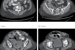
Meanwhile, a deep-learning model used with preoperative CT can predict the complexity and outcomes of abdominal surgery more accurately than surgeons' judgment, according to Dutch research published on 7 July in JAMA Surgery.
Finally, as a bit of much-needed light relief, we bring you the tale of radiologist Dr. Adrian "George" Ringer. He helped bring a lot of new imaging technology to the people of Bermuda, including the island's first MRI system. History expert Dr. Adrian Thomas tells the story of Ringer's unconventional life.


![Overview of the study design. (A) The fully automated deep learning framework was developed to estimate body composition (BC) (defined as subcutaneous adipose tissue [SAT] in liters; visceral adipose tissue [VAT] in liters; skeletal muscle [SM] in liters; SM fat fraction [SMFF] as a percentage; and intramuscular adipose tissue [IMAT] in deciliters) from MRI. The fully automated framework comprised one model (model 1) to quantify different BC measures (SAT, VAT, SM, SMFF, and IMAT) as three-dimensional (3D) measures from whole-body MRI scans. The second model (model 2) was trained to identify standardized anatomic landmarks along the craniocaudal body axis (z coordinate field), which allowed for subdividing the whole-body measures into different subregions typically examined on clinical routine MRI scans (chest, abdomen, and pelvis). (B) BC was quantified from whole-body MRI in over 66,000 individuals from two large population-based cohort studies, the UK Biobank (UKB) (36,317 individuals) and the German National Cohort (NAKO) (30,291 individuals). Bar graphs show age distribution by sex and cohort. BMI = body mass index. (C) After the performance assessment of the fully automated framework, the change in BC measures, distributions, and profiles across age decades were investigated. Age-, sex-, and height-adjusted body composition reference curves were calculated and made publicly available in a web-based z-score calculator (https://circ-ml.github.io).](https://img.auntminnieeurope.com/mindful/smg/workspaces/default/uploads/2026/05/body-comp.XgAjTfPj1W.jpg?auto=format%2Ccompress&fit=crop&h=112&q=70&w=112)


