In a special Christmas edition, a former BMJ editor has reflected on the 20th anniversary of one of the journal's most popular articles: MRI of male and female genitals during coitus and female sexual arousal.
Nobody at the BMJ "thought the study was particularly useful clinically or scientifically, but it contained 'a striking image using a new technology, and everyone agreed that readers might be interested to see it,' " wrote Dr. Tony Delamothe in his new feature (BMJ, December 18, 2019).
 Image of coitus from the famous Dutch study of eight couples imaged by MRI during sexual intercourse. All clinical images courtesy of Dr. Pek van Andel.
The legendary article went on to receive the Ig Nobel Prize for medicine. This dubious honor is given to researchers who explore rather unorthodox, whimsical, and notably less mainstream topics that are designed to both amuse and educate.
The 1999 study by Schultz et al from University Hospital Groningen in the Netherlands did draw reader attention purely based on -- at least one would hope -- the researchers' own observational findings, which were inspired, in part, by a Leonardo da Vinci Renaissance drawing circa 1493.
"The Copulation" prominently depicted a male subject and how da Vinci believed a man would "deliver the goods," so to speak. Da Vinci theorized that a man's sperm traveled from his brain and down his spine. He also surmised that a woman's right lactiferous duct originated in her right breast and ended in the genital area.
"Even a genius like Leonardo da Vinci distorted men's and women's bodies -- as seen now -- to fit the ideology of his time and to the notions of his colleagues, who he paid tribute to," Schultz and colleagues wrote in their study.
To their credit, the Dutch researchers dispelled that misinformation from the Renaissance, as well as the conventional wisdom of the 1960s, as espoused by William Masters and Virginia Johnson, the tandem who received notoriety as researchers of human sexual habits and performance.
Real challenges
As one might expect, there were some serious challenges in getting the Dutch project off the ground back in 1999.
Schultz and colleagues cited "obtrusive and sniffing press hounds," as well as some "problems with sexual performance" among the subjects. Still, in the end, they achieved good images of coitus from 13 "experiments" that included eight couples and three single women.
The determined researchers eventually concluded that MR images of coitus are both "feasible and beautiful." In addition, the shape of a man's penis, as seen on MRI, was not what previous research had suggested, and the size of a woman's uterus did not increase during sexual arousal.
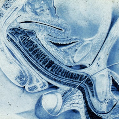
 Midsagittal image of the anatomy of sexual intercourse. P = penis, Ur = urethra, Pe = perineum, U = uterus, S = symphysis, B = bladder, I = intestine, L5 = lumbar 5, Sc = scrotum.
The BMJ paper was hardly the medical equivalent of a moon landing, so why did visitors come flocking in such numbers? Delamothe suggested that the "prospect of seeing coitus on screen (for free) was the attraction, even if all that was on offer was a series of black and white still photographs."
As for the rest of the details, perhaps they are better read in front of a warm fire on a cold winter's night with a heated beverage of choice in hand. Cheers!