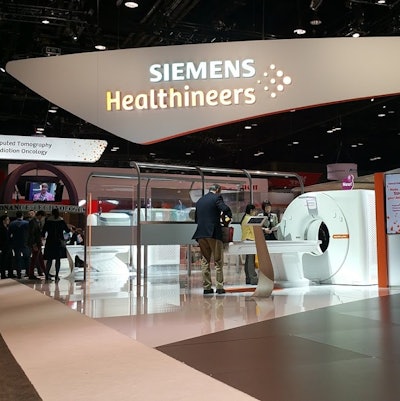
The Siemens Healthineers division of German industrial conglomerate Siemens AG saw revenues remain flat for the fourth quarter of fiscal 2017, but after adjustment for currency fluctuations, the business grew 5%.
For the period, the health business turned in revenue of 3.705 billion euros ($4.31 billion), essentially flat compared with revenue before currency adjustment of 3.698 billion euros ($4.31 billion). However, after currency adjustment, revenue grew 5%.
Operating profit at the health division came in at 702 million euros ($817 million), compared with 696 million euros ($810 million) in the fourth quarter of fiscal 2016. Orders grew 2% on an actual basis and 7% after currency adjustment.
The company said order intake was up due to growth in the diagnostic imaging business and large orders in its service business; growth came mainly in China and Europe and the area representing the Commonwealth of Independent States, Africa, and the Middle East.
For the full year, Siemens Healthineers posted revenue of 13.789 billion euros ($16.05 billion), up 2% on an actual basis and 3% after currency adjustment, compared with sales of 13.535 billion euros ($15.76 billion) for fiscal 2016. The health group's annual operating profit was 2.490 billion euros ($2.90 billion), compared with 2.325 billion euros ($2.71 billion) for fiscal 2016.
In other news, Siemens CEO Joe Kaeser provided additional details about the initial public offering (IPO) of Siemens Healthineers in an interview with CNBC this week.
Siemens previously announced that it plans to spin off the division in an IPO as a way to maximize the value of the unit and enable it to use its own shares to make acquisitions in the healthcare space. While Siemens originally was targeting August 2018 for the IPO, Kaeser told CNBC that it will have completed internal preparations by March of that year, and it will make the move when market conditions are right. Kaeser said the unit was a "fascinating healthcare business."
Finally, Siemens announced on November 9 the appointment of Jochen Schmitz as the new chief financial officer of Siemens Healthineers. Schmitz is currently head of accounting, reporting, and controlling for Siemens; he will succeed Thomas Rathmann, who is leaving Siemens Healthineers by mutual agreement.


![Examples of ultrasound findings and techniques. (A) Images in a 39-year-old male patient with a mass in the left thigh. The mass is heterogeneous on the B-mode US image (compared with the patient in D) and showed increased microvascularity (superb microvascular imaging [SMI]) and shear-wave elastography (SWE) values. Undifferentiated pleomorphic sarcoma was diagnosed at biopsy (with pleomorphic rhabdomyosarcoma in surgical specimen). (B) Images in an 18-year-old male patient with a mass in the left leg. The mass is hypoechoic on the B-mode image, with no other findings suggestive of malignancy. The lesion is in contact with the cortex of the tibia, which is slightly irregular. CT revealed a doubtful anteromedial tibial erosion. The microvascular study demonstrated high vascularization, suggestive of malignancy. Periosteal Ewing sarcoma was diagnosed with both histologic and immunohistochemical confirmation. (C) Images in a 69-year-old female patient with a lump growing on the outside of the left leg. Multiple SWE examinations were performed (please note the high values obtained in the measurements, whereas the color map highlights the stiffness relative to adjacent tissues). SMI showed areas of increased vascularization to target for sampling. Undifferentiated spindle cell sarcoma was diagnosed at biopsy, with residual leiomyosarcoma in the surgical specimen after neoadjuvant therapy. (D) Images in a 56-year-old female patient with a mass in the right thigh. The mass is heterogeneous at both B-mode ultrasound (similar to patient A) and MRI (coronal T2-weighted spectral attenuated inversion recovery [SPAIR]; T1-weighted pre-contrast and postcontrast imaging), which even shows uptake after the administration of paramagnetic contrast material, which is traditionally suggestive of malignancy. Low values at SMI and elastography are suggestive of benignity. Spindle cell lipoma was diagnosed at biopsy, with atypical spindle cell lipomatous tumor in the surgical specimen.](https://img.auntminnieeurope.com/mindful/smg/workspaces/default/uploads/2026/08/images-radiol250278fig2.APCFLSvX6p.jpg?auto=format%2Ccompress&fit=crop&h=100&q=70&w=100)


![Examples of ultrasound findings and techniques. (A) Images in a 39-year-old male patient with a mass in the left thigh. The mass is heterogeneous on the B-mode US image (compared with the patient in D) and showed increased microvascularity (superb microvascular imaging [SMI]) and shear-wave elastography (SWE) values. Undifferentiated pleomorphic sarcoma was diagnosed at biopsy (with pleomorphic rhabdomyosarcoma in surgical specimen). (B) Images in an 18-year-old male patient with a mass in the left leg. The mass is hypoechoic on the B-mode image, with no other findings suggestive of malignancy. The lesion is in contact with the cortex of the tibia, which is slightly irregular. CT revealed a doubtful anteromedial tibial erosion. The microvascular study demonstrated high vascularization, suggestive of malignancy. Periosteal Ewing sarcoma was diagnosed with both histologic and immunohistochemical confirmation. (C) Images in a 69-year-old female patient with a lump growing on the outside of the left leg. Multiple SWE examinations were performed (please note the high values obtained in the measurements, whereas the color map highlights the stiffness relative to adjacent tissues). SMI showed areas of increased vascularization to target for sampling. Undifferentiated spindle cell sarcoma was diagnosed at biopsy, with residual leiomyosarcoma in the surgical specimen after neoadjuvant therapy. (D) Images in a 56-year-old female patient with a mass in the right thigh. The mass is heterogeneous at both B-mode ultrasound (similar to patient A) and MRI (coronal T2-weighted spectral attenuated inversion recovery [SPAIR]; T1-weighted pre-contrast and postcontrast imaging), which even shows uptake after the administration of paramagnetic contrast material, which is traditionally suggestive of malignancy. Low values at SMI and elastography are suggestive of benignity. Spindle cell lipoma was diagnosed at biopsy, with atypical spindle cell lipomatous tumor in the surgical specimen.](https://img.auntminnieeurope.com/mindful/smg/workspaces/default/uploads/2026/08/images-radiol250278fig2.APCFLSvX6p.jpg?auto=format%2Ccompress&fit=crop&h=112&q=70&w=112)