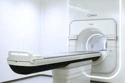
Radiation oncology vendor Elekta said its investigational Unity MR radiation therapy (MR/RT) system is the subject of 21 abstracts at the American Association of Physicists in Medicine (AAPM) annual meeting this week in Denver.
The research includes a presentation from the Clinical Cancer Center at Froedtert & the Medical College of Wisconsin that will share initial results of system-level tests on the MR-linac. The work-in-progress system successfully passed multiple tests assessing imaging and dosimetry performance, and the results also demonstrated successful delivery and measurement of an intensity-modulated radiation therapy (IMRT) treatment plan. Furthermore, the integration of a linear accelerator and high-field MRI did not compromise the performance of either system, Elekta said.
In another talk, researchers from University Medical Center Utrecht in the Netherlands will report that the performance of the MR-linac's experimental software can support real-time applications, according to the company. The group found that the system's real-time motion compensation method software took 62 msec to reconstruct and transport image data after acquisition. In addition, the overall time to gate the beam was 407.6 msec, including the real-time image processing of five images per second, Elekta said.
The company noted that University Medical Center Utrecht used the MR-linac to treat its first patient in May. Other sites in the Elekta MR-Linac Consortium are expected to initiate human trials over the next several months, and Elekta said it remains on track to seek CE Mark approval in the second half of 2017. Elekta plans to file a 510(k) application with the U.S. Food and Drug Administration (FDA) in 2018.