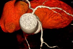
As many as one-fourth of heart attack patients who would normally have a stent implanted can forgo the procedure in favor of conservative management based on the results of optical coherence tomography (OCT), concluded U.S. researchers in a presentation at the European Society of Cardiology congress (ESC 2016) in Rome.
The study used OCT to differentiate plaque erosion from plaque rupture among 405 patients presenting at the emergency department and undergoing coronary angiography, said lead investigator Dr. Ik-Kyung Jang, PhD, from Harvard Medical School in Cambridge, Massachusetts, and Massachusetts General Hospital in Boston.
Distinguishing plaque rupture causes
About 60% of acute coronary syndrome (ACS) cases are caused by plaque rupture, but plaque erosion accounts for another 25% to 44% of cases. These cases have a distinct pathology that is amenable to antithrombotic therapy. OCT can tell the difference between the two.
OCT identified plaque erosion as the underlying pathology in 103 (25.4%) patients. Of these, 60 had a residual diameter stenosis of less than 70% on angiography, a thrombolysis in myocardial infarction (TIMI) flow grade of 3, and were stable without symptoms, the group reported.
These patients were cleared to receive antithrombotic medications alone without stent placement, and were treated with dual antiplatelet therapy (aspirin and ticagrelor), in addition to a glycoprotein IIb/IIIa inhibitor in 63.6%.
At a one-month follow-up, 47 of the 60 patients (78.3%) met the primary endpoint of the study, a greater than 50% reduction in clot size, with 22 patients having no visible clot at all, Jang reported. Overall clot volume decreased from 3.7 mm3 to 0.2 mm3 and minimal flow area increased from 1.7 mm2 to 2.1 mm2.
In the group without stenting, one patient died of gastrointestinal bleeding while on dual antiplatelet therapy; a second patient saw no improvement in the stenotic artery at one month.
Currently all patients with acute coronary syndrome are uniformly treated with stenting regardless of underlying pathology, the researchers said. This study is the first to show that patients with acute coronary syndrome due to erosion may benefit from a noninvasive approach relying on antithrombotic medications, Jang said.
The ability to identify erosion and pursue conservative management may lead to triaging patients away from invasive catheterization, the group concluded.


![Examples of ultrasound findings and techniques. (A) Images in a 39-year-old male patient with a mass in the left thigh. The mass is heterogeneous on the B-mode US image (compared with the patient in D) and showed increased microvascularity (superb microvascular imaging [SMI]) and shear-wave elastography (SWE) values. Undifferentiated pleomorphic sarcoma was diagnosed at biopsy (with pleomorphic rhabdomyosarcoma in surgical specimen). (B) Images in an 18-year-old male patient with a mass in the left leg. The mass is hypoechoic on the B-mode image, with no other findings suggestive of malignancy. The lesion is in contact with the cortex of the tibia, which is slightly irregular. CT revealed a doubtful anteromedial tibial erosion. The microvascular study demonstrated high vascularization, suggestive of malignancy. Periosteal Ewing sarcoma was diagnosed with both histologic and immunohistochemical confirmation. (C) Images in a 69-year-old female patient with a lump growing on the outside of the left leg. Multiple SWE examinations were performed (please note the high values obtained in the measurements, whereas the color map highlights the stiffness relative to adjacent tissues). SMI showed areas of increased vascularization to target for sampling. Undifferentiated spindle cell sarcoma was diagnosed at biopsy, with residual leiomyosarcoma in the surgical specimen after neoadjuvant therapy. (D) Images in a 56-year-old female patient with a mass in the right thigh. The mass is heterogeneous at both B-mode ultrasound (similar to patient A) and MRI (coronal T2-weighted spectral attenuated inversion recovery [SPAIR]; T1-weighted pre-contrast and postcontrast imaging), which even shows uptake after the administration of paramagnetic contrast material, which is traditionally suggestive of malignancy. Low values at SMI and elastography are suggestive of benignity. Spindle cell lipoma was diagnosed at biopsy, with atypical spindle cell lipomatous tumor in the surgical specimen.](https://img.auntminnieeurope.com/mindful/smg/workspaces/default/uploads/2026/08/images-radiol250278fig2.APCFLSvX6p.jpg?auto=format%2Ccompress&fit=crop&h=100&q=70&w=100)


![Examples of ultrasound findings and techniques. (A) Images in a 39-year-old male patient with a mass in the left thigh. The mass is heterogeneous on the B-mode US image (compared with the patient in D) and showed increased microvascularity (superb microvascular imaging [SMI]) and shear-wave elastography (SWE) values. Undifferentiated pleomorphic sarcoma was diagnosed at biopsy (with pleomorphic rhabdomyosarcoma in surgical specimen). (B) Images in an 18-year-old male patient with a mass in the left leg. The mass is hypoechoic on the B-mode image, with no other findings suggestive of malignancy. The lesion is in contact with the cortex of the tibia, which is slightly irregular. CT revealed a doubtful anteromedial tibial erosion. The microvascular study demonstrated high vascularization, suggestive of malignancy. Periosteal Ewing sarcoma was diagnosed with both histologic and immunohistochemical confirmation. (C) Images in a 69-year-old female patient with a lump growing on the outside of the left leg. Multiple SWE examinations were performed (please note the high values obtained in the measurements, whereas the color map highlights the stiffness relative to adjacent tissues). SMI showed areas of increased vascularization to target for sampling. Undifferentiated spindle cell sarcoma was diagnosed at biopsy, with residual leiomyosarcoma in the surgical specimen after neoadjuvant therapy. (D) Images in a 56-year-old female patient with a mass in the right thigh. The mass is heterogeneous at both B-mode ultrasound (similar to patient A) and MRI (coronal T2-weighted spectral attenuated inversion recovery [SPAIR]; T1-weighted pre-contrast and postcontrast imaging), which even shows uptake after the administration of paramagnetic contrast material, which is traditionally suggestive of malignancy. Low values at SMI and elastography are suggestive of benignity. Spindle cell lipoma was diagnosed at biopsy, with atypical spindle cell lipomatous tumor in the surgical specimen.](https://img.auntminnieeurope.com/mindful/smg/workspaces/default/uploads/2026/08/images-radiol250278fig2.APCFLSvX6p.jpg?auto=format%2Ccompress&fit=crop&h=112&q=70&w=112)