Dear MRI Insider,
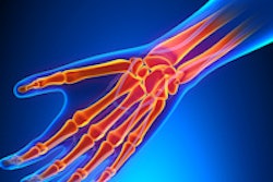
When it comes to the wrist, MRI excels, as shown by two important studies highlighted in your MRI Community.
First up is some original research conducted at the prestigious Heidelberg University in Germany. The group asked whether delayed gadolinium-enhanced MRI of cartilage and T2 mapping are feasible at 3 tesla. To read more, click here.
The second study involved professional golfers, among whom wrist injuries are surprisingly common. In particular, MRI is reliable in documenting the extent of dorsal capsular thickening, as well as any concomitant injuries or bone marrow changes elsewhere in the wrist. Get the story here.
Carrying on the sporting theme, we've posted a news report about the Olympic sport of synchronized swimming and how MRI is contributing to better management of their injuries, particularly of the hip. Click here to learn more.
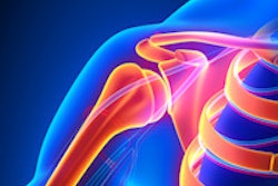
Meanwhile, in shoulder imaging, MRI is playing a valuable role in assessing the bone morphology of the glenoid (version and bone stock) as well as rotator cuff integrity, according to some award-winning work from the U.K. Click here to find out more.
Our recent article about how Lego models can reduce kids' anxiety about MRI has proved incredibly popular, and now the author is planning to exhibit his work at the upcoming RSNA meeting in Chicago. If you missed this report, click here.
This letter features only a few of the many articles posted over recent weeks in the MRI Community. Please do check out the rest of our coverage below this message.


![Overview of the study design. (A) The fully automated deep learning framework was developed to estimate body composition (BC) (defined as subcutaneous adipose tissue [SAT] in liters; visceral adipose tissue [VAT] in liters; skeletal muscle [SM] in liters; SM fat fraction [SMFF] as a percentage; and intramuscular adipose tissue [IMAT] in deciliters) from MRI. The fully automated framework comprised one model (model 1) to quantify different BC measures (SAT, VAT, SM, SMFF, and IMAT) as three-dimensional (3D) measures from whole-body MRI scans. The second model (model 2) was trained to identify standardized anatomic landmarks along the craniocaudal body axis (z coordinate field), which allowed for subdividing the whole-body measures into different subregions typically examined on clinical routine MRI scans (chest, abdomen, and pelvis). (B) BC was quantified from whole-body MRI in over 66,000 individuals from two large population-based cohort studies, the UK Biobank (UKB) (36,317 individuals) and the German National Cohort (NAKO) (30,291 individuals). Bar graphs show age distribution by sex and cohort. BMI = body mass index. (C) After the performance assessment of the fully automated framework, the change in BC measures, distributions, and profiles across age decades were investigated. Age-, sex-, and height-adjusted body composition reference curves were calculated and made publicly available in a web-based z-score calculator (https://circ-ml.github.io).](https://img.auntminnieeurope.com/mindful/smg/workspaces/default/uploads/2026/05/body-comp.XgAjTfPj1W.jpg?auto=format%2Ccompress&fit=crop&h=112&q=70&w=112)


