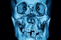
Growth in the conebeam CT (CBCT) segment by far outstrips sales in other dental x-ray segments and almost all other medical imaging modalities in terms of unit growth globally, according to a new report by IHS Medical Technology.
Rising demand for implant dentistry, maxillofacial surgery, and orthodontics are the major factors driving strong market demand for CBCT, the firm said. Globally, the number of CBCT units being shipped is forecast to grow at a compound annual growth rate (CAGR) of 10.8% from 2014 to 2019, generating almost 722 million euros in annual revenue by 2019.
CBCT equipment is replacing extraoral systems such as cephalometric and panoramic systems in all emerging countries. CBCT is seeing an increase in popularity due to its ease of use and also its operational efficiency when compared with other extraoral imaging devices. CBCT offers a view of the dentition that can be used to visualize both erupted and nonerupted teeth, tooth root orientation, and anomalous structures that conventional 2D imaging cannot, according to IHS.
New CBCT systems with three-in-one functionality are being launched that combine CBCT, panoramic, and cephalometric techniques in one system. Planmeca, Danaher, and Sirona are the leading manufacturers in the CBCT market globally, IHS said.
North America was estimated to be the largest regional market for CBCT systems in 2014 with the fastest growth in unit shipments of all the dental x-ray products in the Americas market from 2015 to 2019. This is primarily due to the prevalence of aesthetic dentistry and other more specialized dental services in the U.S. and Canada. Private dental practitioners in this region show the highest adoption rates for advanced dental services, IHS added.
In other markets, such as India and China, uptake of CBCT equipment is still low. Though there has been price erosion in recent years, CBCT is still one of the highest-priced equipment among all other dental x-ray segments. In established markets such as Italy and Japan, CBCT is showing considerable growth by replacing panoramic and cephalometric x-ray systems.
Some of price-sensitive markets in the Asia-Pacific and Europe, Middle East, and Africa regions are upgrading from analog x-ray units to photostimulable phosphor plates and complementary metal oxide semiconductor (CMOS) sensors. These digital sensors are faster and more efficient in generating images with lower radiation risk, according to IHS.


![Examples of ultrasound findings and techniques. (A) Images in a 39-year-old male patient with a mass in the left thigh. The mass is heterogeneous on the B-mode US image (compared with the patient in D) and showed increased microvascularity (superb microvascular imaging [SMI]) and shear-wave elastography (SWE) values. Undifferentiated pleomorphic sarcoma was diagnosed at biopsy (with pleomorphic rhabdomyosarcoma in surgical specimen). (B) Images in an 18-year-old male patient with a mass in the left leg. The mass is hypoechoic on the B-mode image, with no other findings suggestive of malignancy. The lesion is in contact with the cortex of the tibia, which is slightly irregular. CT revealed a doubtful anteromedial tibial erosion. The microvascular study demonstrated high vascularization, suggestive of malignancy. Periosteal Ewing sarcoma was diagnosed with both histologic and immunohistochemical confirmation. (C) Images in a 69-year-old female patient with a lump growing on the outside of the left leg. Multiple SWE examinations were performed (please note the high values obtained in the measurements, whereas the color map highlights the stiffness relative to adjacent tissues). SMI showed areas of increased vascularization to target for sampling. Undifferentiated spindle cell sarcoma was diagnosed at biopsy, with residual leiomyosarcoma in the surgical specimen after neoadjuvant therapy. (D) Images in a 56-year-old female patient with a mass in the right thigh. The mass is heterogeneous at both B-mode ultrasound (similar to patient A) and MRI (coronal T2-weighted spectral attenuated inversion recovery [SPAIR]; T1-weighted pre-contrast and postcontrast imaging), which even shows uptake after the administration of paramagnetic contrast material, which is traditionally suggestive of malignancy. Low values at SMI and elastography are suggestive of benignity. Spindle cell lipoma was diagnosed at biopsy, with atypical spindle cell lipomatous tumor in the surgical specimen.](https://img.auntminnieeurope.com/mindful/smg/workspaces/default/uploads/2026/08/images-radiol250278fig2.APCFLSvX6p.jpg?auto=format%2Ccompress&fit=crop&h=100&q=70&w=100)


![Examples of ultrasound findings and techniques. (A) Images in a 39-year-old male patient with a mass in the left thigh. The mass is heterogeneous on the B-mode US image (compared with the patient in D) and showed increased microvascularity (superb microvascular imaging [SMI]) and shear-wave elastography (SWE) values. Undifferentiated pleomorphic sarcoma was diagnosed at biopsy (with pleomorphic rhabdomyosarcoma in surgical specimen). (B) Images in an 18-year-old male patient with a mass in the left leg. The mass is hypoechoic on the B-mode image, with no other findings suggestive of malignancy. The lesion is in contact with the cortex of the tibia, which is slightly irregular. CT revealed a doubtful anteromedial tibial erosion. The microvascular study demonstrated high vascularization, suggestive of malignancy. Periosteal Ewing sarcoma was diagnosed with both histologic and immunohistochemical confirmation. (C) Images in a 69-year-old female patient with a lump growing on the outside of the left leg. Multiple SWE examinations were performed (please note the high values obtained in the measurements, whereas the color map highlights the stiffness relative to adjacent tissues). SMI showed areas of increased vascularization to target for sampling. Undifferentiated spindle cell sarcoma was diagnosed at biopsy, with residual leiomyosarcoma in the surgical specimen after neoadjuvant therapy. (D) Images in a 56-year-old female patient with a mass in the right thigh. The mass is heterogeneous at both B-mode ultrasound (similar to patient A) and MRI (coronal T2-weighted spectral attenuated inversion recovery [SPAIR]; T1-weighted pre-contrast and postcontrast imaging), which even shows uptake after the administration of paramagnetic contrast material, which is traditionally suggestive of malignancy. Low values at SMI and elastography are suggestive of benignity. Spindle cell lipoma was diagnosed at biopsy, with atypical spindle cell lipomatous tumor in the surgical specimen.](https://img.auntminnieeurope.com/mindful/smg/workspaces/default/uploads/2026/08/images-radiol250278fig2.APCFLSvX6p.jpg?auto=format%2Ccompress&fit=crop&h=112&q=70&w=112)