A team of international researchers have discovered a specific network of brain regions through MRI that is more vulnerable to unhealthy aging, such as Alzheimer's disease, and disorders in young people, such as schizophrenia.
The first-of-its-kind research, published online on 24 November in the Proceedings of the National Academy of Sciences, also concluded that these brain areas in healthy people are the last to develop and the first to show signs of neurodegeneration.
Led by Medical Research Council (MRC) funded researcher Dr. Gwenaëlle Douaud at the Oxford University Functional MRI of the Brain (FMRIB) Center, the study evaluated MR images and patterns in the brain structure of 484 healthy people, ranging in age from 8 to 85.
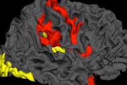
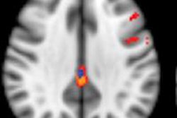
MRI unveiled a network within nerve cells, also known as gray matter, linking mostly "higher order" regions that connect information from different senses that does not develop until late adolescence or early adulthood. That delayed development can affect intellectual ability and long-term memory and later become schizophrenia in young people or Alzheimer's in adults. In addition, this network was found to develop later than the rest of the brain and was the first to degenerate in older age.
"These complex regions, which combine information coming from various senses, seem to be more vulnerable than the rest of the brain to both schizophrenia and Alzheimer's, even though these two diseases have different origins and appear at very different, almost opposite, times of life," the authors noted.
Hugh Perry, PhD, chairman of the MRC's Neurosciences and Mental Health Board, which funded the work, said the findings raise important issues about possible genetic and environmental factors that may occur in early life and then have lifelong consequences.