A new molecular imaging technique that can visualize inflammation in the joints was unveiled at this week's Society of Nuclear Medicine and Molecular Imaging (SNMMI) meeting taking place in St. Louis.
Dutch researchers used PET and SPECT, both of which image physiological processes, with the help of specialized detectors that pick up signals from injected radionuclide imaging agents.
In this case, the researchers evaluated antifibroblast activation protein (FAP) antibodies involved in the inflammation associated with rheumatoid arthritis. They used radiotracers that combine the molecular compound 28H1, which can bind to FAP in the body, with the radionuclides indium-111 (In-111), used in conjunction with SPECT imaging systems, and zirconium (Zr-89), used with PET systems.
The research is novel because radiolabeled anti-FAP antibodies have never been used before in molecular imaging for rheumatoid arthritis, according to lead author Peter Laverman, PhD, assistant professor of nuclear medicine from the department of radiology and nuclear medicine at Radboud University Medical Center in Nijmegen, the Netherlands.
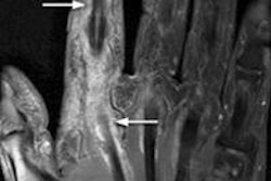
The preclinical study used small animal scanners. Both In-111 28H1 and Zr-89 28H1 showed significantly increased imaging agent uptake in inflamed joints. The uptake was three to four times higher with these agents than another antibody agent evaluated as a control. The researchers also evaluated FDG to image inflammation, but uptake was not associated with the severity of inflammation. This experimental model proved 28H1 tagged with either In-111 or Zr-89 is a superior method for imaging arthritis.
"To the best of our knowledge, high-contrast images of this kind were unheard of until now," Laverman said. He estimated it may take two or more years to accumulate enough research to get the agents approved for arthritis imaging in mainstream clinical practice.