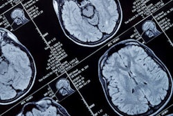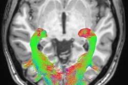
Researchers in Italy used 7-tesla MRI to uncover details of an area in the brain linked to Parkinson's disease, which could help improve early detection of the condition, according to a study published online 26 February in Radiology.
With ultrahigh-field MRI, the group was able to distinguish a three-layered organization of the substantia nigra, a crescent-shaped mass of cells in the midbrain. Based on abnormalities identified in the substantia nigra, the researchers correctly classified patients with Parkinson's disease with a sensitivity of 100% and specificity of 96.2%.
The findings confirm 7-tesla MRI's ability to show the borders of the substantia nigra and its inner organization, as well as radiological signs of change in Parkinson's patients, wrote lead author Dr. Mirco Cosottini, from the University of Pisa, and colleagues.
The MR images "allowed us to discriminate with near-perfect accuracy Parkinson's disease subjects from age-matched healthy subjects," demonstrating 7-tesla MRI's diagnostic power, they wrote (Radiology, 26 February 2014).
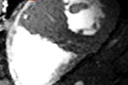
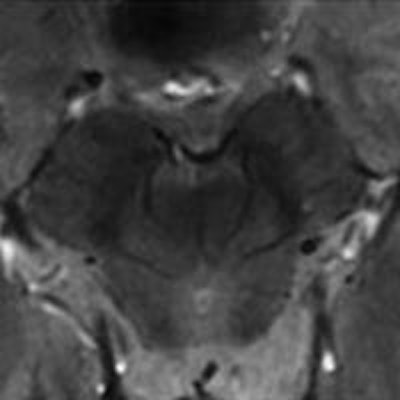
 Seven-tesla MR in vivo images show three levels of the substantia nigra in a 61-year-old male Parkinson's patient. The loss of normal anatomy of the substantia nigra with Parkinson's is characterized by the disappearance of an oval-shaped bright spot in the lateral part of the substantia nigra at level I (left) and by the loss of the hyperintense intermediate layer of the substantia nigra at level II (center). Images courtesy of Radiology.
Parkinson's disease
Parkinson's disease results from the loss of dopamine-producing cells located in the substantia nigra region of the brain. Dopamine is a key neurotransmitter involved in motor and behavioral processes such as mood, reward, addiction, and stress.
Without accurate imaging techniques for diagnosis, clinicians have had to rely on medical history and neurological exams, which can make it challenging to distinguish Parkinson's disease from other neurological ailments.
"Parkinson's disease diagnosis remains clinically based, but with the introduction of 7-tesla MRI into clinical practice, a supporting radiologic diagnosis can be made," Cosottini said in a release from RSNA about the study.
To first assess the normal anatomy of the substantia nigra, the researchers performed both ex vivo and in vivo imaging using a 7-tesla MR system (MR950, GE Healthcare) with a two-channel transmission and 32-channel receiver head coil.
For the ex vivo exam, the group used a substantia nigra specimen from a 67-year-old woman with no adverse neurologic and psychiatric history, who underwent autopsy after a cardiac-related death. The in vivo 7-tesla MRI exams were performed on eight healthy subjects (mean age, 40.1 years).
After training on the appearance of the normal substantia nigra, two blinded neuroradiologists then evaluated 17 subjects with Parkinson's (mean age, 56.9 years) and 13 healthy subjects (mean age, 54.7 years) to determine 7-tesla MRI's ability to differentiate the two groups.
Subjects were considered to have Parkinson's if at least one of three levels of one side of the substantia nigra was abnormal.
Abnormalities
The two readers agreed in their description of a three-layered organization of the substantia nigra, and they were also able to distinguish Parkinson's patients from healthy subjects, the researchers found.
The first reader found abnormalities in at least one of three substantia nigra levels in 17 of 17 Parkinson's patients, while the substantia nigra was normal in 13 of 13 healthy subjects. Sensitivity, specificity, positive predictive value, and negative predictive value for the diagnosis of Parkinson's were each 100%.
The second reader found abnormal areas in 17 of 17 Parkinson's patients and normal results in 12 of 13 healthy subjects. Thus, sensitivity was 100%, specificity was 92%, positive predictive value was 94%, and negative predictive value was 100%.
"In our study, 7-tesla MR targeted images of the midbrain provided a refined depiction of substantia nigra internal organization, which allowed for the identification of three tiers of different signal intensity along the posterior-anterior axis of the midbrain in healthy subjects," the authors wrote.
In addition, Cosottini and colleagues believe the study is the first to demonstrate a "robust MR imaging sign that promptly identifies Parkinson's disease patients with respect to controls," they wrote.
The researchers are also exploring the use of 7-tesla MRI for other neurodegenerative diseases such as mild cognitive impairment.