The combination of functional resection laser surgery and intensity-modulated radiotherapy (IMRT) treatment is more effective at extending the lives of patients with advanced head and neck cancers than having only radiotherapy treatment, according to a study presented at the recent RSNA 2011 conference in Chicago.
Both therapies have been well established as sole treatments for head and neck cancers. To determine if both treatments combined would be more effective, a team of German researchers from Südharz Hospital Nordhausen analyzed the outcomes of 191 of their patients treated between 2001 and 2009. Twenty-six patients, ranging in age from 29 to 92 years, had stage III and 165 patients had stage IV head and neck cancers.
A total of 28 patients were treated for laryngeal cancer, three-fourths of whom received the combined therapies. The remaining 163 patients received treatment for other head and neck cancers, with 76% receiving the combined treatment.
Functional resection laser surgery offered the advantages of preserving more anatomical structures, taking a shorter amount of time to perform, and lowering risks to patients, according to presenter Dr. Jens Buentzel of the university's department of otolaryngology.
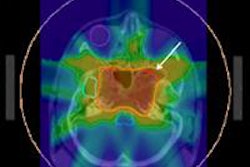
IMRT was used so that organs such as the brain, salivary glands, larynx, and vertebral column could be preserved. Patients received daily fractional treatments of 1.8 Gy, for a total dose of 61.2 Gy for the primary tumor and the N+ neck region and a total dose of 50.4 Gy for the N0 neck region. The median time between surgery and radiotherapy treatment was four weeks.
As part of their analysis, the researchers used data from the tumor registry of the state Thuringia, analyzing only IMRT patients without simultaneous chemotherapy. Patients with laryngeal cancer were separated from other head and neck cancers due to prognostic differences.
Patients were followed for a minimum of 18 months up to 12 years after treatment. Patients with laryngeal cancer who received the combined treatment had a survival rate of 56% at three years, compared with 34% for patients who received only IMRT treatment. Patients with combined therapy also had better survival at five years following treatment (47% versus 34%) and at 10 years following treatment (37% versus 17%). Patients who had the combined treatment lived a median of 3.9 years, compared with 1.6 years for those who had only IMRT.
Patients with other head and neck cancers who had the combined treatment also lived longer: 46% survived three years after treatment, compared with 29%. They also lived more than twice as long, a median of two and a half years, compared with less than a year.
Both groups experienced a similar level of acute toxicities, none of which was higher than stage III. This was very encouraging because the majority of patients were old and had more comorbidities than other cancer patients, Buentzel said. He and his colleagues are currently conducting an evaluation of late toxicities.


![Examples of ultrasound findings and techniques. (A) Images in a 39-year-old male patient with a mass in the left thigh. The mass is heterogeneous on the B-mode US image (compared with the patient in D) and showed increased microvascularity (superb microvascular imaging [SMI]) and shear-wave elastography (SWE) values. Undifferentiated pleomorphic sarcoma was diagnosed at biopsy (with pleomorphic rhabdomyosarcoma in surgical specimen). (B) Images in an 18-year-old male patient with a mass in the left leg. The mass is hypoechoic on the B-mode image, with no other findings suggestive of malignancy. The lesion is in contact with the cortex of the tibia, which is slightly irregular. CT revealed a doubtful anteromedial tibial erosion. The microvascular study demonstrated high vascularization, suggestive of malignancy. Periosteal Ewing sarcoma was diagnosed with both histologic and immunohistochemical confirmation. (C) Images in a 69-year-old female patient with a lump growing on the outside of the left leg. Multiple SWE examinations were performed (please note the high values obtained in the measurements, whereas the color map highlights the stiffness relative to adjacent tissues). SMI showed areas of increased vascularization to target for sampling. Undifferentiated spindle cell sarcoma was diagnosed at biopsy, with residual leiomyosarcoma in the surgical specimen after neoadjuvant therapy. (D) Images in a 56-year-old female patient with a mass in the right thigh. The mass is heterogeneous at both B-mode ultrasound (similar to patient A) and MRI (coronal T2-weighted spectral attenuated inversion recovery [SPAIR]; T1-weighted pre-contrast and postcontrast imaging), which even shows uptake after the administration of paramagnetic contrast material, which is traditionally suggestive of malignancy. Low values at SMI and elastography are suggestive of benignity. Spindle cell lipoma was diagnosed at biopsy, with atypical spindle cell lipomatous tumor in the surgical specimen.](https://img.auntminnieeurope.com/mindful/smg/workspaces/default/uploads/2026/08/images-radiol250278fig2.APCFLSvX6p.jpg?auto=format%2Ccompress&fit=crop&h=100&q=70&w=100)


![Examples of ultrasound findings and techniques. (A) Images in a 39-year-old male patient with a mass in the left thigh. The mass is heterogeneous on the B-mode US image (compared with the patient in D) and showed increased microvascularity (superb microvascular imaging [SMI]) and shear-wave elastography (SWE) values. Undifferentiated pleomorphic sarcoma was diagnosed at biopsy (with pleomorphic rhabdomyosarcoma in surgical specimen). (B) Images in an 18-year-old male patient with a mass in the left leg. The mass is hypoechoic on the B-mode image, with no other findings suggestive of malignancy. The lesion is in contact with the cortex of the tibia, which is slightly irregular. CT revealed a doubtful anteromedial tibial erosion. The microvascular study demonstrated high vascularization, suggestive of malignancy. Periosteal Ewing sarcoma was diagnosed with both histologic and immunohistochemical confirmation. (C) Images in a 69-year-old female patient with a lump growing on the outside of the left leg. Multiple SWE examinations were performed (please note the high values obtained in the measurements, whereas the color map highlights the stiffness relative to adjacent tissues). SMI showed areas of increased vascularization to target for sampling. Undifferentiated spindle cell sarcoma was diagnosed at biopsy, with residual leiomyosarcoma in the surgical specimen after neoadjuvant therapy. (D) Images in a 56-year-old female patient with a mass in the right thigh. The mass is heterogeneous at both B-mode ultrasound (similar to patient A) and MRI (coronal T2-weighted spectral attenuated inversion recovery [SPAIR]; T1-weighted pre-contrast and postcontrast imaging), which even shows uptake after the administration of paramagnetic contrast material, which is traditionally suggestive of malignancy. Low values at SMI and elastography are suggestive of benignity. Spindle cell lipoma was diagnosed at biopsy, with atypical spindle cell lipomatous tumor in the surgical specimen.](https://img.auntminnieeurope.com/mindful/smg/workspaces/default/uploads/2026/08/images-radiol250278fig2.APCFLSvX6p.jpg?auto=format%2Ccompress&fit=crop&h=112&q=70&w=112)