Digital chest tomosynthesis may be a promising first-line tool for lung cancer screening, a preliminary study out of Italy shows. Using the modality with a digital radiography system detected noncalcified nodules in a percentage comparable with CT scans in previous studies.
Low-dose CT is currently the method of choice for lung cancer screening, but due to radiation concerns, researchers are always on the lookout for another option. Digital chest tomosynthesis is a possibility because it creates 3D reconstructions of x-ray images, providing better sensitivity and specificity than conventional radiography. Plus it has a much lower radiation dose than CT.
A total sample size of 2,000 smokers or former smokers of at least 20 pack years is planned for the study, which was presented during a poster session at the RSNA meeting. Subjects must not have shown previous malignancy within five years, said Dr. Maurizio Grosso from Santa Croce e Carle Hospital in Cueno, Italy. Radiologists will perform two digital tomosynthesis scans, one at baseline and one at one-year follow-up. If digital tomosynthesis reveals nodules larger than 5 mm, a low-dose chest CT must be performed.
As of March 2011, 851 subjects had been enrolled. Their mean age was 60, and 68% are currently active smokers. Hypertension, chronic obstructive pulmonary disease, and cardiovascular diseases were the more prevalent comorbidities. Of the digital tomosynthesis exams performed, noncalcified nodules were detected in 28.9% of the cases. Approximately 7% of subjects had nodules larger than 5 mm and thus underwent a CT scan. Lung cancer lesions were detected in 2.6% of subjects, and a three-month follow-up CT scan was prescribed for 0.9% of subjects.
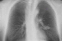
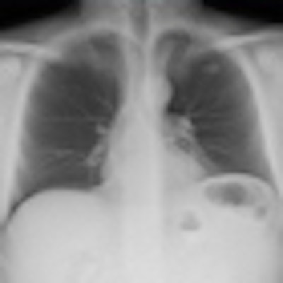
Digital tomosynthesis detected a left apical nodule in a 52-year-old male smoker. All images courtesy of Stephane Chauvie, PhD, and Dr. Maurizio Grosso.
CT confirmed the nodule in the patient and consequently he underwent surgery, which confirmed the tumoral nature of the nodule."We have very preliminary data, but we believe digital tomosynthesis is a quite good method of detection for nodules," Grosso said.
The effective dose for digital tomosynthesis was 0.15 mSv, while the effective dose for CT was 4 mSv.
"It's a balance between image quality and the dose," said Stephane Chauvie, PhD, the medical physicist associated with the study. "For CT you can have good image quality, but you have higher dose. ... This could work as well as a low-dose CT. What has to be found is how much less the dose could be and how if you detect the same nodules, what is the sensitivity, what is the specificity of the technique?"
The other added bonus of using digital tomosynthesis is the cost -- in Italy, it's different but digital tomosynthesis is about 15% of the cost of a CT scanner, Grosso said. A digital tomosynthesis machine costs about $25,000 (19,100 euros) while a CT scanner costs about $300,000 (229,000 euros).
While the results are promising, the researchers caution they are preliminary. In January 2012, they'll be able to present the results of all 2,000 patients, and in January 2013, they can present the data from one year of follow-up.
Two other studies also have shown how promising digital chest tomosynthesis is -- one from Sweden illustrating tomosynthesis' ability to reduce the number of chest CTs, and another from Italy showing its ability to characterize pulmonary lesions.


![Examples of ultrasound findings and techniques. (A) Images in a 39-year-old male patient with a mass in the left thigh. The mass is heterogeneous on the B-mode US image (compared with the patient in D) and showed increased microvascularity (superb microvascular imaging [SMI]) and shear-wave elastography (SWE) values. Undifferentiated pleomorphic sarcoma was diagnosed at biopsy (with pleomorphic rhabdomyosarcoma in surgical specimen). (B) Images in an 18-year-old male patient with a mass in the left leg. The mass is hypoechoic on the B-mode image, with no other findings suggestive of malignancy. The lesion is in contact with the cortex of the tibia, which is slightly irregular. CT revealed a doubtful anteromedial tibial erosion. The microvascular study demonstrated high vascularization, suggestive of malignancy. Periosteal Ewing sarcoma was diagnosed with both histologic and immunohistochemical confirmation. (C) Images in a 69-year-old female patient with a lump growing on the outside of the left leg. Multiple SWE examinations were performed (please note the high values obtained in the measurements, whereas the color map highlights the stiffness relative to adjacent tissues). SMI showed areas of increased vascularization to target for sampling. Undifferentiated spindle cell sarcoma was diagnosed at biopsy, with residual leiomyosarcoma in the surgical specimen after neoadjuvant therapy. (D) Images in a 56-year-old female patient with a mass in the right thigh. The mass is heterogeneous at both B-mode ultrasound (similar to patient A) and MRI (coronal T2-weighted spectral attenuated inversion recovery [SPAIR]; T1-weighted pre-contrast and postcontrast imaging), which even shows uptake after the administration of paramagnetic contrast material, which is traditionally suggestive of malignancy. Low values at SMI and elastography are suggestive of benignity. Spindle cell lipoma was diagnosed at biopsy, with atypical spindle cell lipomatous tumor in the surgical specimen.](https://img.auntminnieeurope.com/mindful/smg/workspaces/default/uploads/2026/08/images-radiol250278fig2.APCFLSvX6p.jpg?auto=format%2Ccompress&fit=crop&h=100&q=70&w=100)


![Examples of ultrasound findings and techniques. (A) Images in a 39-year-old male patient with a mass in the left thigh. The mass is heterogeneous on the B-mode US image (compared with the patient in D) and showed increased microvascularity (superb microvascular imaging [SMI]) and shear-wave elastography (SWE) values. Undifferentiated pleomorphic sarcoma was diagnosed at biopsy (with pleomorphic rhabdomyosarcoma in surgical specimen). (B) Images in an 18-year-old male patient with a mass in the left leg. The mass is hypoechoic on the B-mode image, with no other findings suggestive of malignancy. The lesion is in contact with the cortex of the tibia, which is slightly irregular. CT revealed a doubtful anteromedial tibial erosion. The microvascular study demonstrated high vascularization, suggestive of malignancy. Periosteal Ewing sarcoma was diagnosed with both histologic and immunohistochemical confirmation. (C) Images in a 69-year-old female patient with a lump growing on the outside of the left leg. Multiple SWE examinations were performed (please note the high values obtained in the measurements, whereas the color map highlights the stiffness relative to adjacent tissues). SMI showed areas of increased vascularization to target for sampling. Undifferentiated spindle cell sarcoma was diagnosed at biopsy, with residual leiomyosarcoma in the surgical specimen after neoadjuvant therapy. (D) Images in a 56-year-old female patient with a mass in the right thigh. The mass is heterogeneous at both B-mode ultrasound (similar to patient A) and MRI (coronal T2-weighted spectral attenuated inversion recovery [SPAIR]; T1-weighted pre-contrast and postcontrast imaging), which even shows uptake after the administration of paramagnetic contrast material, which is traditionally suggestive of malignancy. Low values at SMI and elastography are suggestive of benignity. Spindle cell lipoma was diagnosed at biopsy, with atypical spindle cell lipomatous tumor in the surgical specimen.](https://img.auntminnieeurope.com/mindful/smg/workspaces/default/uploads/2026/08/images-radiol250278fig2.APCFLSvX6p.jpg?auto=format%2Ccompress&fit=crop&h=112&q=70&w=112)