Dutch researchers have discovered that hippocampal shape can predict dementia in the general population on average four years before clinical diagnosis. The shape varies from patient to patient, but similarities exist in certain "landmark points," delegates learned at last week's RSNA meeting.
MRI examinations from 511 participants in the Rotterdam Scan study, which started in 1995/1996, were included in the research by Hakim Achterberg, a doctoral student from the department of radiology at Erasmus Medical Center in Rotterdam, the Netherlands, and colleagues. Participants were followed for incidence of dementia using cognitive screening and medical records for 10 years. During that time, 52 subjects developed dementia with an average of four years between the MRI examination and diagnosis.
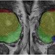
Achterberg and colleagues segmented the left and right hippocampus using an automatic method followed by visual inspection and manual correction in case of errors. In total, 1,024 corresponding landmark points were placed on each hippocampal surface. The shapes were scaled to identical volume so that only shape information, not volume, was contained in the model.
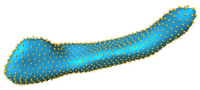 More than a thousand landmark points were placed on the hippocampal surface. All images courtesy of Hakim Achterberg.
The mathematical trick is to link together all these numbers into one big vectpr, he said.
A support vector machine classifier was trained to discriminate between shapes of subjects who stayed cognitively intact during the follow-up and those who developed dementia. Also included in the research was a subset of 50 subjects who developed dementia and 150 age- and gender-matched controls.
The area under the receiver operating characteristic curve was 0.75 when using only shape information, compared with 0.70 when using only volume information. When combining volume and shape the area under the curve increased slightly to 0.76. In the regression model with only volume, the volume term was significant (p < 0.0001). When adding shape, volume was no longer significant (p = 0.1109) and shape was significant (p < 0.0001).
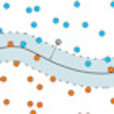
"If you imagine the blue dots being shapes of people who won't develop dementia, the orange dots shapes of people who will develop dementia, and the gray dot a shape of a person whom you don't know whether he will develop dementia, then the gray-blue boundary can be found (in our case using a support vector machine)," Achterberg told AuntMinnieEurope.com. "Once we have this boundary, we check on which side of the boundary this point is. In this case, it is in the side where the blue dots are, so where we estimate the subjects not to develop dementia. The distance from the boundary is how sure we are that the shape really belongs to the estimated group. In this case, we can be reasonably sure the shape indicates the person will not develop dementia."
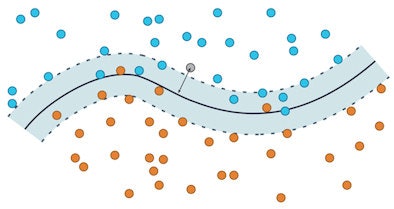 The side of the curve is what determines the estimation whether the subject will develop dementia. The distance from the curve is the certainty the estimation is correction -- the further from the boundary, the more likely the estimation is correct.
Shape is slightly more predictive of dementia, he continued. In the regression model, volume is not any longer significant and shape is more significant.
When asked what the shape is exactly, Achterberg said, "It's very hard to show 'It's this little area shaped like this and that's the big difference.' It's very complicated." There is not a single prototypical shape for healthy or diseased subjects, and there is not a single shape change that indicates dementia, he added.
The researchers also asked whether hippocampal shape can predict dementia before any clinical symptoms.
"This was interesting. We want to look at dementia before people get sick because we might be able to delay it," he said. "Volume becomes less significant, but completely based on shape does surprisingly well. We can still show some predictive value for hippocampal shape."
