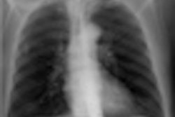
An experimental drug that delivers small doses of radiation to bone metastases in prostate cancer patients has shown that it can prolong survival significantly, according to research presented at the European Multidisciplinary Cancer Congress (EMCC), held in Stockholm this week.
In fact, the phase III clinical trial of the drug radium-223 chloride, also called Alpharadin, was stopped in June 2011 because of the high success rate of the patients randomized to receive the alpha-pharmaceutical in addition to standard treatment.
Patients taking radium-233 chloride had a 30% lower death rate compared with patients taking a placebo, reported Dr. Chris Parker, a consultant clinical oncologist at London's Royal Marsden Hospital. Median survival for this patient cohort, at 14 months, was almost three months longer than the group that received the placebo.
The 922 participants in the Alpharadin in Symptomatic Prostate Cancer (ALSYMPCA) international clinical trial all were resistant to hormone treatment and had bone metastases. The researchers chose to study the drug in patients with prostate cancer because of its high tendency to metastasize to bone. Almost 80% of men with advanced prostate cancer will develop bone metastases.
Alpha-pharmaceuticals work by delivering minute, highly charged, and targeted doses of damaging radiation to bone metastasis. Radium is similar to calcium in that it sticks to bone, particularly to where new bone is being formed, and therefore is a highly efficient means of delivering radiation to a target.
"It takes only a single alpha particle to kill a cell," Parker said. "Collateral damage is minimized because the particles have a tiny range, a few millions of a meter, or a micrometer."
Side effects were reported to be minor. Some patients had nausea and occasional loose bowel movements, but overall, patients tolerated the drug much better than chemotherapy, he noted.
The researchers will now submit their data for regulatory approval to use the drug for treatment of prostate cancer patients. They also hope that additional clinical trials will be conducted, because they believe that the drug could be used to treat many other types of cancer that metastasize to the bone.


![Examples of ultrasound findings and techniques. (A) Images in a 39-year-old male patient with a mass in the left thigh. The mass is heterogeneous on the B-mode US image (compared with the patient in D) and showed increased microvascularity (superb microvascular imaging [SMI]) and shear-wave elastography (SWE) values. Undifferentiated pleomorphic sarcoma was diagnosed at biopsy (with pleomorphic rhabdomyosarcoma in surgical specimen). (B) Images in an 18-year-old male patient with a mass in the left leg. The mass is hypoechoic on the B-mode image, with no other findings suggestive of malignancy. The lesion is in contact with the cortex of the tibia, which is slightly irregular. CT revealed a doubtful anteromedial tibial erosion. The microvascular study demonstrated high vascularization, suggestive of malignancy. Periosteal Ewing sarcoma was diagnosed with both histologic and immunohistochemical confirmation. (C) Images in a 69-year-old female patient with a lump growing on the outside of the left leg. Multiple SWE examinations were performed (please note the high values obtained in the measurements, whereas the color map highlights the stiffness relative to adjacent tissues). SMI showed areas of increased vascularization to target for sampling. Undifferentiated spindle cell sarcoma was diagnosed at biopsy, with residual leiomyosarcoma in the surgical specimen after neoadjuvant therapy. (D) Images in a 56-year-old female patient with a mass in the right thigh. The mass is heterogeneous at both B-mode ultrasound (similar to patient A) and MRI (coronal T2-weighted spectral attenuated inversion recovery [SPAIR]; T1-weighted pre-contrast and postcontrast imaging), which even shows uptake after the administration of paramagnetic contrast material, which is traditionally suggestive of malignancy. Low values at SMI and elastography are suggestive of benignity. Spindle cell lipoma was diagnosed at biopsy, with atypical spindle cell lipomatous tumor in the surgical specimen.](https://img.auntminnieeurope.com/mindful/smg/workspaces/default/uploads/2026/08/images-radiol250278fig2.APCFLSvX6p.jpg?auto=format%2Ccompress&fit=crop&h=100&q=70&w=100)


![Examples of ultrasound findings and techniques. (A) Images in a 39-year-old male patient with a mass in the left thigh. The mass is heterogeneous on the B-mode US image (compared with the patient in D) and showed increased microvascularity (superb microvascular imaging [SMI]) and shear-wave elastography (SWE) values. Undifferentiated pleomorphic sarcoma was diagnosed at biopsy (with pleomorphic rhabdomyosarcoma in surgical specimen). (B) Images in an 18-year-old male patient with a mass in the left leg. The mass is hypoechoic on the B-mode image, with no other findings suggestive of malignancy. The lesion is in contact with the cortex of the tibia, which is slightly irregular. CT revealed a doubtful anteromedial tibial erosion. The microvascular study demonstrated high vascularization, suggestive of malignancy. Periosteal Ewing sarcoma was diagnosed with both histologic and immunohistochemical confirmation. (C) Images in a 69-year-old female patient with a lump growing on the outside of the left leg. Multiple SWE examinations were performed (please note the high values obtained in the measurements, whereas the color map highlights the stiffness relative to adjacent tissues). SMI showed areas of increased vascularization to target for sampling. Undifferentiated spindle cell sarcoma was diagnosed at biopsy, with residual leiomyosarcoma in the surgical specimen after neoadjuvant therapy. (D) Images in a 56-year-old female patient with a mass in the right thigh. The mass is heterogeneous at both B-mode ultrasound (similar to patient A) and MRI (coronal T2-weighted spectral attenuated inversion recovery [SPAIR]; T1-weighted pre-contrast and postcontrast imaging), which even shows uptake after the administration of paramagnetic contrast material, which is traditionally suggestive of malignancy. Low values at SMI and elastography are suggestive of benignity. Spindle cell lipoma was diagnosed at biopsy, with atypical spindle cell lipomatous tumor in the surgical specimen.](https://img.auntminnieeurope.com/mindful/smg/workspaces/default/uploads/2026/08/images-radiol250278fig2.APCFLSvX6p.jpg?auto=format%2Ccompress&fit=crop&h=112&q=70&w=112)