Researchers from several institutions believe they have achieved a breakthrough in MRI that would enable brain scans to be performed more than seven times faster than currently possible.
 |
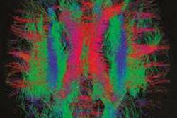
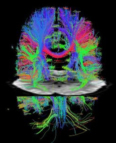
| Colored tracks show the direction of nerve fiber bundles, providing a 3D image of the axonal pathways in the white matter (cortex) of a resting human brain. The entire 3D image was scanned in 8.5 minutes instead of 30 minutes. Image courtesy of David Feinberg. |
The lead author of the study is David Feinberg, a physicist and adjunct professor in UCB's Helen Wills Neuroscience Institute and president of the company Advanced MRI Technologies in Sebastopol, CA.
Feinberg noted that because the technique works on all modern MRI scanners, the impact of the ultrafast imaging technique will be immediate and widespread at research institutions worldwide.
In addition to broadly advancing the field of neuroimaging, the discovery will have immediate effects on the Human Connectome Project, funded last year by the National Institutes of Health (NIH) to map the connections of the human brain through functional MRI (fMRI) and structural MRI scans of 1,200 healthy adults.
The faster scans are made possible by combining two technical improvements invented in the past decade that separately boosted scanning speeds two to four times more than what was already the fastest MRI technique, echo planar imaging.
Physical limitations of each method prevented further speed improvements, but together their image accelerations are multiplied, Feinberg said.
Related Reading
Functional MRI helps detect effects of nicotine on brain, January 4, 2011
Functional MRI may track aberrant pediatric brain development, September 13, 2010
Study offers strategy for managing incidental findings on fMRI, July 6, 2010
Resting fMRI exam may predict stroke and brain injury outcomes, March 30, 2010
fMRI helps find schizophrenia symptoms, January 20, 2009
Copyright © 2011 AuntMinnie.com