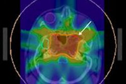
In a U.K. study evaluating dry mouth toxicities, intensity-modulated radiation therapy (IMRT) proved to be far less debilitating for cancer patients than conventional radiation therapy. Results were published online January 12 in Lancet Oncology.
The randomized phase III PARSPORT trial included six U.K. cancer treatment centers participating from 2003 through 2007. The study was designed to determine if reduced irradiation of the parotid glands using IMRT could lower the incidence of severe xerostomia in patients with squamous cell carcinoma of the head and neck, compared to treatment with conventional radiation therapy.
Investigators reported that at a median follow-up of 44 months, the percentage of patients with grade 2 or higher xerostomia side effects was significantly lower in the group of patients who received IMRT.
A total of 94 patients were enrolled in clinical trial: 47 patients for each treatment arm. All patients received 65 Gy in 30 daily fractions delivered over a six-week period. More than 80% in the total group were experiencing grade 2 or higher xerostomia three months after treatment. However, this toxicity had declined significantly one and two years after treatment for the IMRT group.
Cases of xerostomia by treatment modality
|
Principal investigator Christopher Nutting, MD, a consultant clinical oncologist and head of the head and neck unit of the Royal Marsden Hospitals NHS Foundation Trust in London, and colleagues also recorded salivary flow in 80 of the patients prior to treatment. Fifty-nine patients were evaluated one year following treatment, and almost half of the 34 patients in the IMRT group had unstimulated saliva flow from the contralateral parotid gland. However, none of the 25 patients in the conventional radiation therapy group experienced unstimulated saliva flow.
Thirty-one patients were evaluated two years after treatment. Again, none of the patients in the conventional treatment group, a total of 15, had unstimulated saliva flow, whereas seven (44%) of 16 evaluated IMRT patients did.
Patients in the IMRT group experienced more fatigue than their counterparts, a side effect attributed to greater radiation dose to nontumor tissues. At two years, no significant differences were seen between the two groups in nonxerostomia late toxicities, locoregional control, or overall survival, the research team reported.
By Cynthia E. Keen
AuntMinnie.com staff writer
January 18, 2011
Related Reading
IMRT reduces dry mouth toxicity, June 2, 2010
Adverse effects common with chemoradiation for head and neck cancer, December 22, 2009
Acupuncture associated with neuronal activation and physiological responses, August 28, 2008
Copyright © 2011 AuntMinnie.com


![Examples of ultrasound findings and techniques. (A) Images in a 39-year-old male patient with a mass in the left thigh. The mass is heterogeneous on the B-mode US image (compared with the patient in D) and showed increased microvascularity (superb microvascular imaging [SMI]) and shear-wave elastography (SWE) values. Undifferentiated pleomorphic sarcoma was diagnosed at biopsy (with pleomorphic rhabdomyosarcoma in surgical specimen). (B) Images in an 18-year-old male patient with a mass in the left leg. The mass is hypoechoic on the B-mode image, with no other findings suggestive of malignancy. The lesion is in contact with the cortex of the tibia, which is slightly irregular. CT revealed a doubtful anteromedial tibial erosion. The microvascular study demonstrated high vascularization, suggestive of malignancy. Periosteal Ewing sarcoma was diagnosed with both histologic and immunohistochemical confirmation. (C) Images in a 69-year-old female patient with a lump growing on the outside of the left leg. Multiple SWE examinations were performed (please note the high values obtained in the measurements, whereas the color map highlights the stiffness relative to adjacent tissues). SMI showed areas of increased vascularization to target for sampling. Undifferentiated spindle cell sarcoma was diagnosed at biopsy, with residual leiomyosarcoma in the surgical specimen after neoadjuvant therapy. (D) Images in a 56-year-old female patient with a mass in the right thigh. The mass is heterogeneous at both B-mode ultrasound (similar to patient A) and MRI (coronal T2-weighted spectral attenuated inversion recovery [SPAIR]; T1-weighted pre-contrast and postcontrast imaging), which even shows uptake after the administration of paramagnetic contrast material, which is traditionally suggestive of malignancy. Low values at SMI and elastography are suggestive of benignity. Spindle cell lipoma was diagnosed at biopsy, with atypical spindle cell lipomatous tumor in the surgical specimen.](https://img.auntminnieeurope.com/mindful/smg/workspaces/default/uploads/2026/08/images-radiol250278fig2.APCFLSvX6p.jpg?auto=format%2Ccompress&fit=crop&h=100&q=70&w=100)


![Examples of ultrasound findings and techniques. (A) Images in a 39-year-old male patient with a mass in the left thigh. The mass is heterogeneous on the B-mode US image (compared with the patient in D) and showed increased microvascularity (superb microvascular imaging [SMI]) and shear-wave elastography (SWE) values. Undifferentiated pleomorphic sarcoma was diagnosed at biopsy (with pleomorphic rhabdomyosarcoma in surgical specimen). (B) Images in an 18-year-old male patient with a mass in the left leg. The mass is hypoechoic on the B-mode image, with no other findings suggestive of malignancy. The lesion is in contact with the cortex of the tibia, which is slightly irregular. CT revealed a doubtful anteromedial tibial erosion. The microvascular study demonstrated high vascularization, suggestive of malignancy. Periosteal Ewing sarcoma was diagnosed with both histologic and immunohistochemical confirmation. (C) Images in a 69-year-old female patient with a lump growing on the outside of the left leg. Multiple SWE examinations were performed (please note the high values obtained in the measurements, whereas the color map highlights the stiffness relative to adjacent tissues). SMI showed areas of increased vascularization to target for sampling. Undifferentiated spindle cell sarcoma was diagnosed at biopsy, with residual leiomyosarcoma in the surgical specimen after neoadjuvant therapy. (D) Images in a 56-year-old female patient with a mass in the right thigh. The mass is heterogeneous at both B-mode ultrasound (similar to patient A) and MRI (coronal T2-weighted spectral attenuated inversion recovery [SPAIR]; T1-weighted pre-contrast and postcontrast imaging), which even shows uptake after the administration of paramagnetic contrast material, which is traditionally suggestive of malignancy. Low values at SMI and elastography are suggestive of benignity. Spindle cell lipoma was diagnosed at biopsy, with atypical spindle cell lipomatous tumor in the surgical specimen.](https://img.auntminnieeurope.com/mindful/smg/workspaces/default/uploads/2026/08/images-radiol250278fig2.APCFLSvX6p.jpg?auto=format%2Ccompress&fit=crop&h=112&q=70&w=112)