Acupuncture can relieve the symptoms of xerostomia, chronic dry mouth, according to the findings of a randomized clinical trial published online on 23 October in Annals of Oncology. This may be welcome news to the nearly half million patients worldwide diagnosed each year with head and neck cancers.
Xerostomia is a common side effect of radiotherapy treatment, which usually ends within 18 months following treatment. However, up to 41% of patients may have symptoms for five years. The condition can interfere with taste, chewing, swallowing, speaking, and sleeping.
A number of clinical trials have shown that acupuncture can reduce and even may prevent xerostomia. Now another study funded by Cancer Research U.K. supports this.
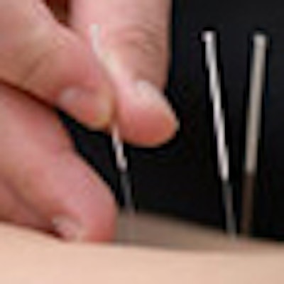
Seven cancer centers in the U.K. enrolled 145 patients suffering from radiation-induced xerostomia. Half of the group received 20-minute acupuncture sessions on a weekly basis for eight weeks; the other half received two one-hour oral care educational sessions, one each month. At the end of eight weeks, each group received the other treatment.
The primary objective of the clinical trial was to measure patient-reported changes in the severity of dry mouth. Secondary objectives were to measure other key xerostomia symptoms, such as changes in saliva production, dry lips, sticky saliva, and the need to sip water to eat or to moisten the mouth.
Although xerostomia symptoms were not alleviated, when patients completed their acupuncture treatments, a higher percentage reported reduction of the condition than those who initially received only education about oral care.
The study did not follow patients to determine if symptoms intensified at three months following treatment, lead author Dr. Richard Simcock, of the department of oncology at Sussex Cancer Centre in Brighton, and co-authors reported. They suggested that additional studies be undertaken to establish the duration of benefit and the length of treatment.
"This is a very neglected group of patients suffering from a most unpleasant side effect of treatment for which all other ameliorative interventions have failed to address adequately. The acupuncture intervention has been designed in a way that allows it to be delivered simply and cheaply in normal hospital surroundings and yet still produces a significant benefit for patients with a chronic symptom," Simcock said.


![Examples of ultrasound findings and techniques. (A) Images in a 39-year-old male patient with a mass in the left thigh. The mass is heterogeneous on the B-mode US image (compared with the patient in D) and showed increased microvascularity (superb microvascular imaging [SMI]) and shear-wave elastography (SWE) values. Undifferentiated pleomorphic sarcoma was diagnosed at biopsy (with pleomorphic rhabdomyosarcoma in surgical specimen). (B) Images in an 18-year-old male patient with a mass in the left leg. The mass is hypoechoic on the B-mode image, with no other findings suggestive of malignancy. The lesion is in contact with the cortex of the tibia, which is slightly irregular. CT revealed a doubtful anteromedial tibial erosion. The microvascular study demonstrated high vascularization, suggestive of malignancy. Periosteal Ewing sarcoma was diagnosed with both histologic and immunohistochemical confirmation. (C) Images in a 69-year-old female patient with a lump growing on the outside of the left leg. Multiple SWE examinations were performed (please note the high values obtained in the measurements, whereas the color map highlights the stiffness relative to adjacent tissues). SMI showed areas of increased vascularization to target for sampling. Undifferentiated spindle cell sarcoma was diagnosed at biopsy, with residual leiomyosarcoma in the surgical specimen after neoadjuvant therapy. (D) Images in a 56-year-old female patient with a mass in the right thigh. The mass is heterogeneous at both B-mode ultrasound (similar to patient A) and MRI (coronal T2-weighted spectral attenuated inversion recovery [SPAIR]; T1-weighted pre-contrast and postcontrast imaging), which even shows uptake after the administration of paramagnetic contrast material, which is traditionally suggestive of malignancy. Low values at SMI and elastography are suggestive of benignity. Spindle cell lipoma was diagnosed at biopsy, with atypical spindle cell lipomatous tumor in the surgical specimen.](https://img.auntminnieeurope.com/mindful/smg/workspaces/default/uploads/2026/08/images-radiol250278fig2.APCFLSvX6p.jpg?auto=format%2Ccompress&fit=crop&h=100&q=70&w=100)


![Examples of ultrasound findings and techniques. (A) Images in a 39-year-old male patient with a mass in the left thigh. The mass is heterogeneous on the B-mode US image (compared with the patient in D) and showed increased microvascularity (superb microvascular imaging [SMI]) and shear-wave elastography (SWE) values. Undifferentiated pleomorphic sarcoma was diagnosed at biopsy (with pleomorphic rhabdomyosarcoma in surgical specimen). (B) Images in an 18-year-old male patient with a mass in the left leg. The mass is hypoechoic on the B-mode image, with no other findings suggestive of malignancy. The lesion is in contact with the cortex of the tibia, which is slightly irregular. CT revealed a doubtful anteromedial tibial erosion. The microvascular study demonstrated high vascularization, suggestive of malignancy. Periosteal Ewing sarcoma was diagnosed with both histologic and immunohistochemical confirmation. (C) Images in a 69-year-old female patient with a lump growing on the outside of the left leg. Multiple SWE examinations were performed (please note the high values obtained in the measurements, whereas the color map highlights the stiffness relative to adjacent tissues). SMI showed areas of increased vascularization to target for sampling. Undifferentiated spindle cell sarcoma was diagnosed at biopsy, with residual leiomyosarcoma in the surgical specimen after neoadjuvant therapy. (D) Images in a 56-year-old female patient with a mass in the right thigh. The mass is heterogeneous at both B-mode ultrasound (similar to patient A) and MRI (coronal T2-weighted spectral attenuated inversion recovery [SPAIR]; T1-weighted pre-contrast and postcontrast imaging), which even shows uptake after the administration of paramagnetic contrast material, which is traditionally suggestive of malignancy. Low values at SMI and elastography are suggestive of benignity. Spindle cell lipoma was diagnosed at biopsy, with atypical spindle cell lipomatous tumor in the surgical specimen.](https://img.auntminnieeurope.com/mindful/smg/workspaces/default/uploads/2026/08/images-radiol250278fig2.APCFLSvX6p.jpg?auto=format%2Ccompress&fit=crop&h=112&q=70&w=112)