When it comes to imaging breast implants for rupture, MRI and ultrasound are recognized widely as the modalities of choice, but a new study by German researchers could change all that because it suggests CT may be a viable alternative.
Just like nonsurgically enhanced women, patients with breast implants also require evaluation of their breast health. Following the scandal involving French manufacturer Poly Implant Prothèse (PIP), who used low-grade industrial silicone for most of their implants, there has been an uptick in the demand for imaging women with augmented breasts to check whether their implants have ruptured.
There are two kinds of ruptures: intracapsular and extracapsular. The rupture depends on the location of the escaped silicone with respect to the fibrous capsule that is built up around the implant. MRI and ultrasound are generally used because silicone implants are too radiodense on mammography. However, MRI is contraindicated in some patients (such as those with pacemakers, cochlear implants, etc.) or not possible because of severe claustrophobia.
"MRI is obviously the technique of choice but is time-consuming and expensive, and may yield a substantial number of false-positive results in a cancer screening setting," wrote Dr. Thorsten R. C. Johnson, from the department of clinical radiology at the University of Munich-Grosshadern Campus in Munich, and colleagues in a study appearing online on 13 October in European Radiology. "So far, CT has not played a role in the evaluation of the breast or implants."
Dual-energy CT has been clinically available since 2006 and offers the possibility of acquiring two CT datasets at the same time using different x-ray spectra, the researchers noted. The two datasets can be analyzed voxel by voxel using a three-material decomposition algorithm to identify materials with photoelectric effect such as silicon.
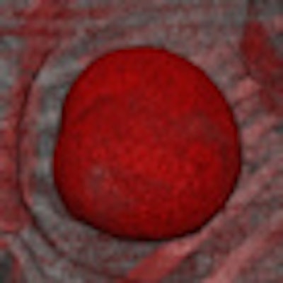
 CT images of a patient with Poly Implant Prothèse (PIP) implants on both sides. The silicone signal is encoded in red. There is a small leak on the medial dorsal aspect of the left implant. The enlarged left axillary lymph nodes show a strong silicone signal. Top: Lateral oblique view. Bottom: Frontal view. All images courtesy of Dr. Thorsten R. C. Johnson.
"I think the remarkable aspect is that we are actually doing sort of molecular imaging in CT -- we are tracing a specific element which represents the pathogenic substrate for disease, similar to [dual-energy] CT for kidney stones or gout," Johnson said in an interview with AuntMinnieEurope.com. "Nobody would have thought this is possible in CT some eight years ago. So we are on a way to specific imaging and characterization of disease rather than purely morphological imaging. I've been involved in the development for dual-energy CT right from the start and it's fascinating to see it thriving."
The researchers examined seven silicone breast implants on dual-source CT (Somatom Definition Flash, Siemens Healthcare) at 100- and 140-kV tube potential with a 0.8-mm tin filter. After the technical feasibility had been confirmed, two patients were included. The silicone of the implant specimens showed a strong dual-energy signal, according to the researchers. In one patient, both implants were intact, while a rupture was identified in the other patient. Ultrasound, MRI, surgical findings, and histology confirmed the dual-energy CT diagnosis.
"Based on our initial results, it seems that dual-energy CT might become an option for implant evaluation in particular cases, especially if there are contraindications for MRI," Johnson and colleagues noted. "Of course, in contrast to emerging high-resolution breast CT, the technique is not sufficient to simultaneously screen for cancer and implies exposure to ionizing radiation."
The equivalent dose of 2.2 and 2.7 mSv in the study was well below the reference limit for chest examinations (DLP 400 mGy*cm). In conventional mammography, patients with implants receive higher radiation doses because of the density of the implants, sometimes 10.7 mGy per breast or a total equivalent dose of 2.6 mSv, so the overall dose is comparable, he added.
"Thus, weighing the health risks of a ruptured implant against the low risk of radiation-induced cancer, the indication may appear similar as in many other diseases that are routinely evaluated by CT," the researchers wrote. "A strength of the technique is the specific depiction of silicone without the administration of contrast material, so there are no other side effects or risks."
Dual-source CT showed a strong silicone signal and indicated implant rupture and silicone in the axillary lymph nodes in one of the patients, which seems to justify the use of this technique.
Several colleagues emailed Johnson to say they thought it was remarkable dual-energy CT works so well, and they think the technique is interesting and promising, Johnson said during his interview.
"Dual-energy CT offers the possibility of specifically visualizing silicone, making it feasible to evaluate breast implants for rupture," the researchers concluded in their study. "A systematic clinical trial will be required to determine the diagnostic accuracy of this technique and to define appropriate indications."
The researchers are preparing further studies on patients who need their implants evaluated and in whom MRI is contraindicated, but they haven't started recruiting yet, Johnson told AuntMinnieEurope.com.
|
Study disclosure One of the co-authors is an employee of Siemens Healthcare. |