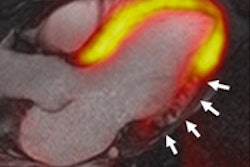
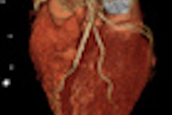
Following up on the CORE 64 study that tested the value of coronary CT angiography vis-à-vis angiography, researchers have launched a new international trial to compare 320-detector-row CT perfusion to SPECT myocardial perfusion imaging (MPI) for detecting early coronary artery disease.
Cardiologists and radiologists at 15 medical centers in eight countries, led by Dr. João Lima and colleagues at Johns Hopkins University in Baltimore, said they have enrolled the first dozen patients in a year-long investigation to determine if perfusion CT can outperform SPECT and other diagnostic tests in tracking the earliest signs of coronary artery stenosis.
Trial participants, all referred for invasive angiography for symptoms such as shortness of breath, chest pain, or fatigue, will also undergo SPECT MPI and 320-detector-row CT (Aquilion One, Toshiba America Medical Systems, Tustin, CA) perfusion imaging.
"Our study goal is to figure out how well various imaging tests measure the degree of blockage or narrowing in any particular artery and therefore which is more useful in predicting patients who need catheterization or angioplasty, or bypass surgery," Lima said in a statement. "Some patients would do just as well or better with drug therapy to maintain a healthy blood flow to the heart, but we need to better sort out who they are with more accuracy."
CT perfusion is fast and exposes patients to less radiation than SPECT, investigators said. If performed correctly, 320-detector-row CT involves an average radiation exposure of about 8 mSv, compared to 10-26 mSv for SPECT and 2-10 mSv for invasive angiography.
Participating CORE 320 sites include Beth Israel Deaconess Medical Center and Brigham and Women's Hospital in Boston; the U.S. National Heart, Lung, and Blood Institute in Bethesda, MD; the University of Toronto in Canada; the University of Copenhagen in Denmark; Charité Medical University in Berlin; Iwate Medical University in Morioka, Japan, and two other Japanese centers; and universities in Singapore and Brazil.
By Eric Barnes
By AuntMinnie.com staff writers
February 10, 2010
Related Reading
Stress perfusion CT adds value to coronary CT angiography, February 9, 2010
Contrast dose halved in coronary CTA, December 2, 2009
DECT cuts tests as a one-stop myocardial, coronary artery exam, August 10, 2009
Adenosine stress DECT equivalent to SPECT, MRI, November 21, 2008
Stress MPI with Tc-99m SPECT identifies high-risk obese patients, September 1, 2006
Copyright © 2010 AuntMinnie.com


![Examples of ultrasound findings and techniques. (A) Images in a 39-year-old male patient with a mass in the left thigh. The mass is heterogeneous on the B-mode US image (compared with the patient in D) and showed increased microvascularity (superb microvascular imaging [SMI]) and shear-wave elastography (SWE) values. Undifferentiated pleomorphic sarcoma was diagnosed at biopsy (with pleomorphic rhabdomyosarcoma in surgical specimen). (B) Images in an 18-year-old male patient with a mass in the left leg. The mass is hypoechoic on the B-mode image, with no other findings suggestive of malignancy. The lesion is in contact with the cortex of the tibia, which is slightly irregular. CT revealed a doubtful anteromedial tibial erosion. The microvascular study demonstrated high vascularization, suggestive of malignancy. Periosteal Ewing sarcoma was diagnosed with both histologic and immunohistochemical confirmation. (C) Images in a 69-year-old female patient with a lump growing on the outside of the left leg. Multiple SWE examinations were performed (please note the high values obtained in the measurements, whereas the color map highlights the stiffness relative to adjacent tissues). SMI showed areas of increased vascularization to target for sampling. Undifferentiated spindle cell sarcoma was diagnosed at biopsy, with residual leiomyosarcoma in the surgical specimen after neoadjuvant therapy. (D) Images in a 56-year-old female patient with a mass in the right thigh. The mass is heterogeneous at both B-mode ultrasound (similar to patient A) and MRI (coronal T2-weighted spectral attenuated inversion recovery [SPAIR]; T1-weighted pre-contrast and postcontrast imaging), which even shows uptake after the administration of paramagnetic contrast material, which is traditionally suggestive of malignancy. Low values at SMI and elastography are suggestive of benignity. Spindle cell lipoma was diagnosed at biopsy, with atypical spindle cell lipomatous tumor in the surgical specimen.](https://img.auntminnieeurope.com/mindful/smg/workspaces/default/uploads/2026/08/images-radiol250278fig2.APCFLSvX6p.jpg?auto=format%2Ccompress&fit=crop&h=100&q=70&w=100)


![Examples of ultrasound findings and techniques. (A) Images in a 39-year-old male patient with a mass in the left thigh. The mass is heterogeneous on the B-mode US image (compared with the patient in D) and showed increased microvascularity (superb microvascular imaging [SMI]) and shear-wave elastography (SWE) values. Undifferentiated pleomorphic sarcoma was diagnosed at biopsy (with pleomorphic rhabdomyosarcoma in surgical specimen). (B) Images in an 18-year-old male patient with a mass in the left leg. The mass is hypoechoic on the B-mode image, with no other findings suggestive of malignancy. The lesion is in contact with the cortex of the tibia, which is slightly irregular. CT revealed a doubtful anteromedial tibial erosion. The microvascular study demonstrated high vascularization, suggestive of malignancy. Periosteal Ewing sarcoma was diagnosed with both histologic and immunohistochemical confirmation. (C) Images in a 69-year-old female patient with a lump growing on the outside of the left leg. Multiple SWE examinations were performed (please note the high values obtained in the measurements, whereas the color map highlights the stiffness relative to adjacent tissues). SMI showed areas of increased vascularization to target for sampling. Undifferentiated spindle cell sarcoma was diagnosed at biopsy, with residual leiomyosarcoma in the surgical specimen after neoadjuvant therapy. (D) Images in a 56-year-old female patient with a mass in the right thigh. The mass is heterogeneous at both B-mode ultrasound (similar to patient A) and MRI (coronal T2-weighted spectral attenuated inversion recovery [SPAIR]; T1-weighted pre-contrast and postcontrast imaging), which even shows uptake after the administration of paramagnetic contrast material, which is traditionally suggestive of malignancy. Low values at SMI and elastography are suggestive of benignity. Spindle cell lipoma was diagnosed at biopsy, with atypical spindle cell lipomatous tumor in the surgical specimen.](https://img.auntminnieeurope.com/mindful/smg/workspaces/default/uploads/2026/08/images-radiol250278fig2.APCFLSvX6p.jpg?auto=format%2Ccompress&fit=crop&h=112&q=70&w=112)