

Dynamic PET imaging moves beyond traditional tumor detection by capturing how radioactive tracers travel through blood vessels in real-time, allowing clinicians to assess both pathology and organ function simultaneously with a single injection.
- Beyond tumor detection: Dynamic PET follows tracer movement through circulation to reveal vascular abnormalities, organ dysfunction, and blood flow patterns invisible to conventional delayed imaging.
- One injection, two types of information: Early dynamic imaging captures both metabolic pathology and vascular physiology, eliminating the need for additional contrast agents.
- Clinical applications: Dynamic PET is used for prostate cancer recurrence, breast cancer, sarcoma, pulmonary emboli detection, and assessment of cardiac transit times and renal blood flow.
- Practical advantages: Particularly valuable for patients with contrast allergies or impaired kidney function, offering vascular information without iodinated contrast agents.
- Implementation challenges: Wider adoption requires advances in data processing, storage capacity, and workflow integration into routine clinical practice.
For decades, PET has been remarkably good at finding tumors. But some of the most valuable information may emerge in the first minutes of a scan, long before the images clinicians usually examine are acquired.
"Whole-body PET created what I like to call lumpology," said Prof. Rodney Hicks of the University of Melbourne, Australia, during the opening session of CHILI 9.0, organized by the European Society for Hybrid, Molecular and Translational Imaging (ESHIMT).
Unlike conventional delayed imaging, dynamic PET follows radioactive tracers as they travel through blood vessels, reach organs, and accumulate in tissue. This allows clinicians to assess not only tumors but also circulation and organ function.
"We can simultaneously assess both pathology and physiology," Hicks said.
What the first minutes reveal
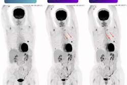
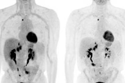
In one patient, dynamic imaging revealed wedge-shaped perfusion defects consistent with multiple pulmonary emboli. In another, a tumor obstructing the superior vena cava forced blood through collateral vessels. Other scans showed asymmetric kidney perfusion, tricuspid regurgitation, and abnormal pulmonary blood flow.
At Hicks' center, dynamic imaging is already part of routine practice for selected indications. Since installing a Biograph Vision Quadra scanner (Siemens Healthineers) in January 2023, the team has performed approximately 15,000 PET studies. Dynamic acquisitions are used in cases including biochemical recurrence of prostate cancer, high-grade breast cancer, sarcoma, ocular melanoma, hepatocellular carcinoma, and head and neck cancer.
"I don't think many people are using it clinically," Hicks said. "We've moved very much into using it clinically."
The technique may be particularly useful for patients with contrast allergies or impaired renal function, offering vascular information without an additional iodinated contrast agent.
One injection, two kinds of information
Prof. Lalit Sundar of Ludwig Maximilian University of Munich, Germany, talked about what happens in the first five minutes after tracer injection. While conventional PET focuses on where a tracer eventually accumulates, early dynamic imaging captures its passage through the circulation, much like a contrast agent.
"With just one injection, you basically have two meanings," Sundar said. Sundar presented examples of pulmonary perfusion abnormalities, an iliac arterial obstruction, and an aortic aneurysm that were not apparent on delayed PET images but became visible when early tracer dynamics were analyzed.
The approach could also provide insights into cardiac transit times and renal blood flow. However, Sundar cautioned that these findings remain preliminary. He described them as "trust me, bro" observations, acknowledging that they have not yet been clinically validated against established imaging methods such as contrast-enhanced CT or ventilation/perfusion imaging.
Some early dynamic analyses may also be feasible on conventional PET scanners with shorter axial fields of view, particularly for organ-specific assessments such as pulmonary perfusion.
Bringing dynamic PET into routine practice
Despite these promising applications, wider clinical adoption faces practical hurdles, including data processing, storage requirements, and integration into existing workflows.
At Hicks' center, approximately 25 patients are scanned daily, but early and delayed acquisitions result in around 40 patient placements on the scanner table. Retaining the raw dynamic data also requires substantial storage capacity.
For Hicks and Sundar, the opportunity is to move beyond locating disease to understanding how it affects the body. Whether the first five minutes of PET will become a routine part of clinical decision-making will depend on further validation and the ability to integrate these additional measurements into everyday practice.