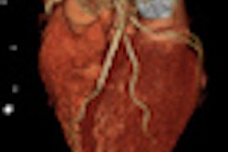
A two-step heart test consisting of 320-detector-row CT angiography (CTA) followed by CT perfusion (CTP) helps separate patients who need invasive angiography from those who do not, according to results of the international CORE 320 study presented on Monday at the European Society of Cardiology (ESC) annual meeting in Munich.
Researchers from 16 centers in eight countries found that CTA's ability to detect flow-limiting coronary artery disease was significantly improved with the addition of 320-detector-row CT perfusion. Compared with invasive angiography as the reference standard, diagnostic accuracy (expressed as area under the receiver operator characteristics curve, or AUC) rose significantly -- from 0.81 with CTA alone to 0.87 using CTA plus CTP. In addition, single-beat cardiac imaging helped keep radiation dose low.
The trial is the culmination of research begun in 2004 that proved the accuracy of perfusion CT imaging in preclinical studies, then in single-center studies, and finally in this multicenter study of 381 patients using 320-detector-row CT technology, said co-author Dr. Richard George, an assistant professor of medicine at Johns Hopkins University.
George said he initiated the research in 2004 and went on to develop the 320-detector-row CTP technique with colleague and co-investigator Dr. João Lima, professor of medicine and radiology and director of cardiovascular imaging at Johns Hopkins. The principal investigator was Dr. Carlos Rochitte from University of São Paulo Medical School.
"What this study really shows is that you're not only able to measure anatomic stenosis with CTA, but also the significance of those stenoses -- and be able to actually see if those stenoses impede blood flow," George told AuntMinnieEurope.com. "We've learned from studies that measure the hemodynamic significance of stenoses that we only want to be treating those patients with coronary stenting or bypass surgery who actually have stenoses that reduce myocardial blood flow, so the 320 study shows us that CT is capable of doing this."
About 30% of patients undergoing catheterization are found to have minimal disease or no blockage, he said. CT perfusion helps clarify the need for invasive angiography in patients with chest pain who do not have evidence of myocardial infarction.
Thanks to coronary CTA's high negative predictive value, patients who are negative at CTA can safely go home, he said. But the course of action is murkier for patients with a positive result on CTA, who may or may not need intervention. Perfusion CT is suitable for these patients.
SPECT myocardial perfusion imaging is the most common noninvasive test for demonstrating reduced blood flow to the heart, but unlike CTP it doesn't indicate the number or specific location of stenoses. The combination of 320-detector-row CTA and CTP enabled the CORE 320 researchers to visualize the anatomy of the stenoses and determine whether they were causing the perfusion deficit, identifying the patients who need revascularization, George said.
Typically, only about 5% to 10% of patients undergoing CTA for chest pain would end up needing a perfusion study, George said, but the CORE 320 study cohort was at higher risk. "The population we used this test for were patients who were already being referred for invasive angiography, so they're patients who have a much higher pretest likelihood of having coronary disease" -- perhaps as high as 40% to 50%. Even at that level, CTP "would give you a chance to still avoid an invasive test," he said.
The researchers performed both CTA and CTP in all 381 patients using 320-detector-row CT (Aquilion One, Toshiba Medical Systems), in addition to angiography and SPECT. The study's primary goals were to determine the accuracy of integrated CTA-CTP for identifying patients with flow-limiting coronary artery disease, defined as having a stenosis of 50% or greater with a corresponding perfusion deficit on stress SPECT myocardial perfusion imaging. All imaging data were examined by independent core laboratories rather than the individual sites, George said.
On a per-patient basis, the diagnostic accuracy of CTA-CTP expressed as AUC for detecting or ruling out flow-limiting coronary artery disease was 0.87 (95% confidence interval [CI]: 0.83-0.91) for 50% or greater stenosis and 0.89 (95% CI: 0.86-0.93) for 70% or greater stenosis. The prevalence of obstructive coronary artery disease using invasive angiography plus SPECT was 38%, compared with 59% for invasive angiography alone, the group reported.
For identifying flow-limiting coronary artery disease, adding CT perfusion boosted accuracy from an AUC of 0.81 (95% CI: 0.77-0.86) for CTA alone to an AUC of 0.87 (95% CI: 0.83-0.91) using integrated CTA-CTP (p < 0.001).
"In a reference standard that's looking at stenosis as well as ischemia caused by that stenosis ... this is excellent diagnostic accuracy," George said. "If you use CTA alone to predict the reference standard, the AUC is 0.81, so when we add in CTP and go to 0.87, the difference is statistically significant. There will be secondary papers that look at the accuracy of SPECT versus CTP, etc.," and more results will be presented later, he said. "But I can tell you they look good," he added.
The wide-volume 320-detector-row CT scanner also helped by minimizing artifact and keeping radiation doses low, limiting scan times to a single heartbeat for CTA and to two beats for CTP in most scans. The median radiation dose for CTA was 3.16 mSv, with an additional (median) 5.31 mSv for the CTP test, George said. Contrast dose was also minimized at 320-detector-row CT, with 81% of patients receiving a total 60 cc of contrast media split between two injections, one for each of the two scans.
Several single-center studies have shown that CT perfusion can be performed accurately on 64-detector-row CT as well, but the 320-detector-row scanner brings several unique advantages to the table, George said. These include the lack of artifacts caused by the need to stitch together smaller volumes from several heartbeats on 64-slice scanners; using multiple heartbeats also impairs contrast uniformity and requires higher contrast doses compared to 320, he said.
The results of the CORE 320 study are also notable considering that many centers were just learning how to use the technology, he said.
"A few sites had experience with CT perfusion, but the majority of our sites had never even done a perfusion study," he said. "In this multicenter international trial, we were able to show that we could perform CT angiography and CT perfusion accurately, with a low radiation dose, and that it predicts very accurately a reference standard currently used in clinical practice, which includes invasive coronary angiogram and a SPECT perfusion study," he said.


