German researchers used 64-slice CT to examine a bust of Nefertiti, discovering new details on the process used to make the sculpture of one of Egypt's most intriguing personalities.
Nefertiti, the wife of the Egyptian pharaoh Akhenaten, was the most renowned great royal wife of all 31 Egyptian dynasties. A bust of her likeness was created 3,300 years ago; discovered in 1912, it is now part of the collection of the Egyptian Museum of Berlin.
CT has been used previously to examine the bust, but not until now have researchers used thin-section MDCT, according to a study published in the April issue of Radiology. The lead author on the study was Dr. Alexander Huppertz, director of the Imaging Science Institute in Berlin.
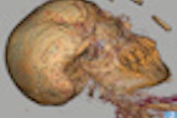
Huppertz and colleagues used 64-slice spiral CT with submillimeter section thickness to examine the bust, assess its conservation status, gain information on its creation, and provide a 3D surface reformation of the bust's inner limestone sculpture.
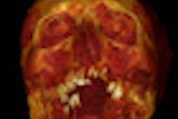
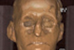
The results showed that a multistep process was used to create the sculpture. The stucco layer on the bust's face and ears is very thin, but the rear part of the reconstructed crown contains two thick stucco layers. CT images showed several fissures and nonuniform bonding between the layers, according to the researchers.
Thin-section CT also provided detailed images of the bust's inner structure and showed the limestone core to be not just a mold, but a skillfully rendered work of art, the authors said. Retouching the creases in the corners of the mouth and smoothing the bump on the nose on the outer face may have been the artist's choice and reflective of the aesthetic ideals of that era, the article said.
Related Reading
Philips CT images Egyptian mummy, February 5, 2009
CT digs into dinosaur's cranial anatomy, feeding habits, December 19, 2007
Cold case closed: CT solves iceman's cause of death, July 4, 2007
Scan artist: Radiologist uses CT to reveal mystery of antiquities, October 25, 2005
CT helps unwrap mummy mystery, March 29, 2005
Copyright © 2009 AuntMinnie.com