NEW YORK (Reuters Health), Sep 3 - Esophageal dilatation visible on a high-resolution CT scan of the lungs may be a sign of scleroderma, according to findings published in the September issue of the Annals of the Rheumatic Diseases.
There is evidence that immunosuppressive treatment in the early stages of systemic sclerosis (SSc) may improve survival, "enhancing the need for early diagnosis and regular evaluation of organ involvement," wrote Dr. M. C. Vonk, of Radboud University Nijmegen Medical Center, the Netherlands, and colleagues.
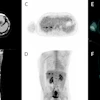
The researchers examined the predictive value of esophageal dilatation on the high-resolution CT (HRCT) scan for the diagnosis of SSc. Included in the study were 105 consecutive patients with scleroderma and 107 consecutive control subjects. Two independent radiologists, who were blinded for the diagnosis of the patients, evaluated the scans for esophageal dilatation and interstitial lung disease.
Infra-aortic esophageal dilatation was observed in 62% of SSc patients and 12% of controls. The positive predictive value of esophageal dilatation for the diagnosis of SSc was 83% and the negative predictive value was 69%.
There was no difference observed in the prevalence of esophageal dilatation in patients with early disease and those with a longer duration of disease. No correlation was found between esophageal dilatation and interstitial lung disease in cases and controls.
"The presence of an esophageal dilatation on an HRCT scan does not completely predict the presence of SSc, and its absence does not rule out SSc," Vonk's team writes. "However, it could be worthwhile if a radiologist, on noticing an esophageal dilatation on an HRCT scan of the chest, reports this finding and perhaps even suggests the possibility of a diagnosis of SSc, enabling the referring specialist to start further diagnostic workup for SSc."
Ann Rheum Dis 2008;67:1317-1321.
Last Updated: 2008-09-02 17:35:44 -0400 (Reuters Health)
Copyright © 2008 Reuters Limited. All rights reserved. Republication or redistribution of Reuters content, including by framing or similar means, is expressly prohibited without the prior written consent of Reuters. Reuters shall not be liable for any errors or delays in the content, or for any actions taken in reliance thereon. Reuters and the Reuters sphere logo are registered trademarks and trademarks of the Reuters group of companies around the world.