Researchers from U.K. artificial intelligence (AI) software developer Lucida Medical and Hampshire Hospitals NHS Foundation Trust have launched a retrospective cohort validation study of the firm's Pi AI-based software for prostate cancer detection on MRI.
The study will include deidentified data from 2,100 patients who have been diagnosed with prostate cancer. The researchers will collaborate to check the performance of the software and, if necessary, calibrate its settings, validate its expected performance, and test it, Lucida said.
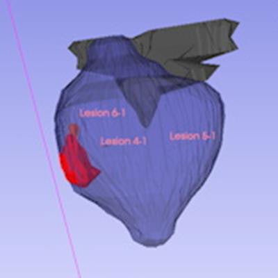
 The figure above illustrates the RTSTRUCT format segmentations output by the software. The segmentations are overlaid on a 3D multiplanar reconstruction of the T2 axial image, together with the 3D mesh view.
A little over half of the data will be utilized to ensure that the software works well across the range of scanners and scanning protocols utilized in different hospitals. This data will be used to check that the software is correctly calibrated and to change settings if needed, according to the vendor. The AI software will then be tested on the rest of the data to assess its performance on MRI exams it hasn't seen yet.
The company said it will publish the data from the study once it's completed.