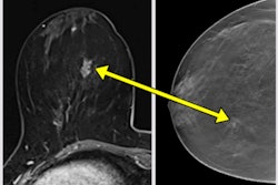
Keep a watchful eye on digital breast tomosynthesis-guided biopsies in 2024. That’s the opinion of researchers in Madrid, who are convinced that these biopsies are fast becoming a more attractive option. Don’t miss our report from RSNA 2023.
Have you ever heard of the Mickey Mouse, picket fence, tattoo, comet, and other signs in breast imaging? Getting to know them can aid interpretation, cut errors, and avoid unnecessary investigations, according to award-winning research also presented recently at the Chicago meeting.
Earlier this week, we unveiled our top 10 list of most viewed articles in 2023. Medicolegal investigations and workforce-related topics were prominent, as they probably will be again next year.
Nearly four years have passed since the first case of COVID-19 was identified, but new studies on the impact of COVID-19 vaccination continue to be published. Check out this latest study from Graz in Austria.
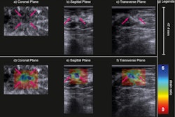
In other news, researchers from Radboud University Medical Center in Nijmegen, the Netherlands, have found that strain elastography can differentiate between malignant and benign lesions when applied to an automated breast volume scanner. Don’t miss our article.
This is our final newsletter for 2023. On behalf of the entire editorial and publishing team, I wish you all the very best for the holidays. We have greatly appreciated your continued support and participation in AuntMinnieEurope.com, and we very much look forward to continuing to serve the radiological community in 2024.
Philip Ward
Editor in Chief





![A normal mammogram confirmed by three-year radiologic follow-up illustrates reader-marked regions of interest (ROIs) during (A) unaided (round 1) and (B) artificial intelligence (AI)–assisted (round 2) reading. Each colored dot represents an ROI for recall by a human reader. Readers could mark more than one ROI per case, represented by multiple dots of the same color. During AI-assisted reading, the AI system displayed three visible prompts: two with suspicion of malignancy scores of 35% (left mediolateral oblique [L MLO] and craniocaudal [L CC]) and one with a suspicion of malignancy score of 10% (right craniocaudal [R CC]), shown as polygonal overlays. Without AI, six of 10 readers (60%) marked a false-positive ROI. With AI assistance, this fell to two of 10 (20%). R MLO = right mediolateral oblique.](https://img.auntminnieeurope.com/mindful/smg/workspaces/default/uploads/2026/07/2026-07-14-radiology-mammogram-ai-auto-bias.H0bYO8QlWs.jpg?auto=format%2Ccompress&fit=crop&h=100&q=70&w=100)


![A normal mammogram confirmed by three-year radiologic follow-up illustrates reader-marked regions of interest (ROIs) during (A) unaided (round 1) and (B) artificial intelligence (AI)–assisted (round 2) reading. Each colored dot represents an ROI for recall by a human reader. Readers could mark more than one ROI per case, represented by multiple dots of the same color. During AI-assisted reading, the AI system displayed three visible prompts: two with suspicion of malignancy scores of 35% (left mediolateral oblique [L MLO] and craniocaudal [L CC]) and one with a suspicion of malignancy score of 10% (right craniocaudal [R CC]), shown as polygonal overlays. Without AI, six of 10 readers (60%) marked a false-positive ROI. With AI assistance, this fell to two of 10 (20%). R MLO = right mediolateral oblique.](https://img.auntminnieeurope.com/mindful/smg/workspaces/default/uploads/2026/07/2026-07-14-radiology-mammogram-ai-auto-bias.H0bYO8QlWs.jpg?auto=format%2Ccompress&fit=crop&h=112&q=70&w=112)