A form of tinnitus that may indicate serious vascular issues can be successfully diagnosed and treated in many cases, thanks to advances in neuroradiological diagnostics and treatment, according to a statement from the German Society for Neuroradiology (DGNR).
Pulse-synchronous tinnitus, which is typically one-sided and follows one’s heartbeat, affects up to 5% of patients with severe tinnitus. The cause of pulse-synchronous tinnitus is often changes in the blood vessels in the head or neck -- e.g., fistulas, vascular constrictions, or malformations -- which may prove dangerous should they obstruct blood flow from the brain.
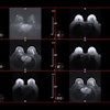
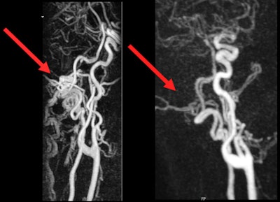 (Left) MRI before intervention. Pathological flow signal in the area of the left occipital condyle can be seen. (Right) MRI after intervention. No more pathological flow signal, and the tinnitus is visible.Images courtesy of UKE Hamburg-Eppendorf and Dr. Fabian Flottmann.
"Patients should be particularly vigilant if the ringing in the ears changes, headaches occur or even neurological deficits such as visual disturbances or dizziness are added," said Dr. Fabian Flottmann, neuroradiologist at the University Medical Center Hamburg-Eppendorf (UKE).
It can be diagnosed using techniques such as contrast-enhanced MRI, time-resolved MR angiography, and CT or catheter angiography to precisely localize vascular changes. In many cases, it can then be causally treated, he explained.
"If we identify the cause, there is a very good chance of recovery," Flottmann said.
Procedures for pulse-synchronous tinnitus are usually minimally invasive and performed via the wrist artery or the inguinal artery. Fistulas may be treated with embolizations, in which the "short circuit" between the artery and vein is closed with platinum coils or tissue adhesive; constrictions in venous drainage pathways may be widened using stents.
Previously, pulse-synchronous tinnitus was more likely to be the purview of specialist areas such as ear, nose, and throat medicine or neurology; however, neuroradiology is increasingly playing a key role due to advances in imaging and minimally invasive therapeutic procedures.
Along with advances in technology, the collaboration between the areas of otolaryngology, neurology, and neuroradiology is critical for the successful diagnosis and therapy for pulse-synchronous tinnitus. Flottmann said that specialist lectures for ear, nose and throat doctors are already held regularly at the UKE, and colleagues in private practice actively seek exchanges.
More details are available here.


