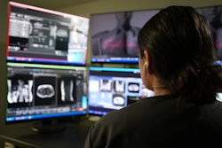
The deployment of deep learning reconstruction (DLR) for MRI at a Finnish academic hospital led to gains in productivity and cost-effectiveness with overall high image quality, according to a recent analysis published on 23 February in the European Journal of Radiology.
A team headed by medical physicist Mikael Brix, PhD, of Oulu University Hospital, found that the optimization reduced MRI scanning time, even with one fewer scanner available than before the DLR implementation.
"[Our] results demonstrate the utility of DLR in improving MRI productivity and support the predictive accuracy of simulation-based health technology assessment," the group wrote.
The need for MR imaging can be met in several ways: procuring new scanners, expanding appointment slots beyond normal working hours, outsourcing part of the service, or increasing the throughput of individual scanners. Brix and colleagues investigated this last option via a study that explored the effect of DLR on MRI workflow in two time periods (preintervention, January to October 2023, and postintervention, January to October 2025).
Preintervention, there were six MRI scanners in the unit; during DLR adoption, there were five, due to a merger and relocation of the pediatric and emergency imaging departments. In fact, it was this reduction in the MRI fleet that provided the impetus to optimize the scanners with DLR, the group noted.
The team analyzed scanner log data for both study time periods, assessed exam and sequence durations, and compared the changes observed with any predicted capacity increases. Four radiologists assessed the quality of the resulting images on a three-point scale to identify any DLR shortcomings using four criteria: noise, image sharpness, artifacts, and diagnostic suitability.
The researchers reported the following results after the DLR was deployed:
- Mean reduction in total exam sequence time for the five scanners was 6.6 minutes, for a total mean reduction of 16%.
- Total exam time decreased by a mean of 4.7 minutes.
- Musculoskeletal exams had the most significant reductions in scan and exam times.
The authors noted variability in these reduction numbers among the individual scanners for a variety of reasons, including a later deployment of DLR on one scanner; a significant increase in the duration of scans and exams in another scanner, likely due to its shift in usage from pediatric and elective patients in pre-DLR period to use as the primary emergency scanner in the DLR implementation period.
As for image quality, the four radiologists deemed the DLR-optimized 2025 scans better overall in the areas of noise reduction and sharpness, while finding “no significant differences” between the 2023 and 2025 images for artifacts or diagnostic suitability.
 Representative artifacts frequently encountered with accelerated deep-learning reconstruction (DLR) sequences, illustrated using images from a healthy volunteer with written informed consent. (Gray arrow) Severe artifacts rendering sagittal spinal cord T2 DLR images nondiagnostic. Evaluation of the sagittal spinal cord T2 DLR images is limited by severe artifacts. (Black arrow) Linear artifacts are enhanced in coronal T2 DLR image. (White arrow) Axial brain image showing signal heterogeneity in the thalamus. In addition, there is blurring in the basal ganglia and insular region.European Journal of Radiology
However, while DLR showed excellent performance for musculoskeletal and prostate imaging in terms of quality, it had limitations in some other areas, especially neurological imaging. The researchers cautioned that DLR seems to amplify motion- and flow-related artifacts, which made it less suited for brain imaging. Additionally, the DLR used was not clinically applicable for the high-resolution 3D sequences commonly used in neurological studies. The authors also found that there was suboptimal image quality in contrast-enhanced studies; as a result, they noted that “DLR sequences have not been approved for contrast-enhanced acquisitions.”
Even given these limitations, the team concluded that DLR’s positive contribution to MRI scanning was considerable: their findings showed the hospital is on track to save approximately €400,000 annually with the DLR deployment (once capacity is fully compensated) compared to operating an additional scanner.
Read the full analysis here.