Implementing a few key interventions can reduce exam delays for MRI exams performed on patients under sedation or general anesthesia, researchers have reported.
A team led by Dr. Aric Lee, of National University Hospital in Singapore found that relatively straightforward initiatives such as workflow standardization and preadmission patient counseling cut delays for these types of exams by more than 30%. The results were published on 18 June in the Journal of the American College of Radiology.
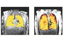
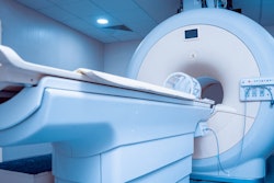
MRI exam delays are common, due to longer scan times the modality requires and safety concerns. What's more, MRI scans conducted under sedation or general anesthesia are even more likely to be delayed, as they require "more complex logistics and coordination of multiple staff members across different hospital departments," the group noted.
"Delays in MRIs, particularly those performed under sedation or general anesthesia, can significantly impact resource utilization, timely diagnosis, and patient satisfaction," the authors wrote.
Lee and colleagues sought to cut delays in MRI exams conducted on patients under sedation or general anesthesia at Singapore's National University Hospital by 15% over a six-month time frame. (The hospital performs 32,000 MRI exams per year.)
The group -- which included radiologists, MRI technologists, radiology and outpatient clinic nursing staff, anesthesiologists, and a pediatrician -- identified particular stages in the imaging process where delays could occur. Lee and colleagues then developed a protocol called "Plan, Do, Study, Act," implementing it over four cycles that included the following four interventions:
- Workflow standardization (for example, exam admission booking)
- Preadmission patient counseling consisting of a single-page checklist
- Clarifying scan times (scheduled time as the start of the scan rather than when the patient should arrive and an effort to ensure patients were in the MRI suite 30 minutes before)
- Written consent for patient transfers from hospital rooms to the MRI suite
For their analysis, they assessed 627 MRI exams performed on patients under sedation or general anesthesia between January 2022 and June 2023. Of these, 88.7% were conducted under sedation, 3.5% under monitored anesthesia, and 7.8% under general anesthesia; 443 were done before the interventions were implemented and 184 after the interventions were put into place. Preintervention, 71.6% of the scans were delayed more than 15 minutes and 28.2% by more than 60 minutes, with a median delay of 30 minutes.
The team did note a trend of increased delay times after intervention one, but this trend shifted to a steady decrease after interventions two, three, and four.
| Delay times for MRI exams performed on patients under sedation or general anesthesia | ||||
|---|---|---|---|---|
| Measure (median) | Baseline | Intervention 1 | Intervention 2 | Intervention 3/4 |
| Delay time (in minutes) | 30 | 40 | 15 | 15 |
| Scans delayed over 15 minutes | 71.6% | 73.7% | 43.5% | 36.9% |
| Scans delayed by 60 minutes or more | 28.2% | 34.2% | 14.5% | 10.7% |
Overall, the interventions resulted in a 34.7%-point reduction in scans delayed over 15 minutes, a 17.5%-point reduction in scans delayed by 60 or more minutes, and a reduced median delay time by 15 minutes, Lee and colleagues reported.
The study findings are promising, but more work needs to be done, according to the authors.
"Unavoidable reasons for delays, such as patient factors (for example, delirious or uncooperative patients, scanners accommodating emergent MRIs) [may] persist," the group concluded. "Other methods of increasing the impact of our interventions, such as integrating the workflow into our hospital electronic medical records and educating patients, are possible future strategies. Further work could also explore the adaptability of our quality improvement interventions across different imaging modalities or in diverse healthcare settings, such as regional hospitals within our network, to further enhance healthcare delivery."
The complete study can be found here.