Dear AuntMinnieEurope Member,
When a radiology department has a mobile MRI scanner onsite that's operated by an outside contractor, the relationship between the two parties can be a gray area. Uncertainty may surround the specific responsibilities of each group, particularly with regard to staff training, and the national regulations may be unclear too. If things go seriously wrong, where does the blame lie?
The question of responsibility has arisen in the case of October's MRI accident in Swedish Lapland. It will be fascinating to see how the police investigation handles this matter. Find out more in the MRI Community.
Dutch researchers have investigated the impact of quality assurance on radiographer reporting, looking closely at nearly half a million examinations from the national breast cancer screening program. They published their findings in Radiology on 7 January. Go to the Women's Imaging Community.
Dr. Mukund Joshi (1942 to 2020) was a charming, modest man and a dedicated teacher. I saw this firsthand at the 1998 International Congress of Radiology in Delhi, India, where he spent the entire conference giving lectures on ultrasound and chatting at length with his huge band of admirers. Nothing pleased him more than passing on his clinical knowledge and inspiring others. Don't miss this tribute to one of radiology's gentlemen.
Also in our Ultrasound Community you can read a fascinating report about a surprise medical discovery by an astronaut. Bizarre things can happen in outer space ...
German investigators have found that dual-energy CT can boost sensitivity and detection in cases of acute pulmonary embolism, particularly for endoluminal clots in small segmental or subsegmental lung vessels. A team from Würzburg has shared its experiences and two sets of striking clinical images in an article posted in the CT Community.
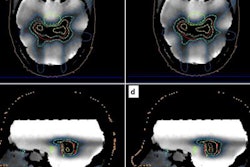
Finally, a group from Athens has elaborated on its use of MR-guided radiotherapy to facilitate the direct visualization of structures of interest in the treatment position, with high soft-tissue contrast. This opens up the possibility of adaptive replanning to compensate for changes in the patient's anatomy or position.


![Overview of the study design. (A) The fully automated deep learning framework was developed to estimate body composition (BC) (defined as subcutaneous adipose tissue [SAT] in liters; visceral adipose tissue [VAT] in liters; skeletal muscle [SM] in liters; SM fat fraction [SMFF] as a percentage; and intramuscular adipose tissue [IMAT] in deciliters) from MRI. The fully automated framework comprised one model (model 1) to quantify different BC measures (SAT, VAT, SM, SMFF, and IMAT) as three-dimensional (3D) measures from whole-body MRI scans. The second model (model 2) was trained to identify standardized anatomic landmarks along the craniocaudal body axis (z coordinate field), which allowed for subdividing the whole-body measures into different subregions typically examined on clinical routine MRI scans (chest, abdomen, and pelvis). (B) BC was quantified from whole-body MRI in over 66,000 individuals from two large population-based cohort studies, the UK Biobank (UKB) (36,317 individuals) and the German National Cohort (NAKO) (30,291 individuals). Bar graphs show age distribution by sex and cohort. BMI = body mass index. (C) After the performance assessment of the fully automated framework, the change in BC measures, distributions, and profiles across age decades were investigated. Age-, sex-, and height-adjusted body composition reference curves were calculated and made publicly available in a web-based z-score calculator (https://circ-ml.github.io).](https://img.auntminnieeurope.com/mindful/smg/workspaces/default/uploads/2026/05/body-comp.XgAjTfPj1W.jpg?auto=format%2Ccompress&fit=crop&h=100&q=70&w=100)


![Overview of the study design. (A) The fully automated deep learning framework was developed to estimate body composition (BC) (defined as subcutaneous adipose tissue [SAT] in liters; visceral adipose tissue [VAT] in liters; skeletal muscle [SM] in liters; SM fat fraction [SMFF] as a percentage; and intramuscular adipose tissue [IMAT] in deciliters) from MRI. The fully automated framework comprised one model (model 1) to quantify different BC measures (SAT, VAT, SM, SMFF, and IMAT) as three-dimensional (3D) measures from whole-body MRI scans. The second model (model 2) was trained to identify standardized anatomic landmarks along the craniocaudal body axis (z coordinate field), which allowed for subdividing the whole-body measures into different subregions typically examined on clinical routine MRI scans (chest, abdomen, and pelvis). (B) BC was quantified from whole-body MRI in over 66,000 individuals from two large population-based cohort studies, the UK Biobank (UKB) (36,317 individuals) and the German National Cohort (NAKO) (30,291 individuals). Bar graphs show age distribution by sex and cohort. BMI = body mass index. (C) After the performance assessment of the fully automated framework, the change in BC measures, distributions, and profiles across age decades were investigated. Age-, sex-, and height-adjusted body composition reference curves were calculated and made publicly available in a web-based z-score calculator (https://circ-ml.github.io).](https://img.auntminnieeurope.com/mindful/smg/workspaces/default/uploads/2026/05/body-comp.XgAjTfPj1W.jpg?auto=format%2Ccompress&fit=crop&h=112&q=70&w=112)


