

Selecting the right imaging approach remains essential to detecting cancer and avoiding unnecessary examinations, experts noted at the Conference on Hybrid Imaging Live (CHILI 9.0).
During a session on precision oncology, speakers discussed what PET, MRI, and diagnostic CT can add to cancer staging and treatment planning. Their examples ranged from tracer-negative lesions to conflicting imaging findings in prostate cancer.
Is low-dose CT enough?
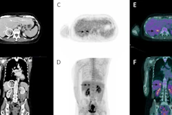
While low-dose CT primarily serves attenuation correction and anatomical localization, diagnostic CT can provide additional information, particularly when performed with intravenous contrast, according to Dr. Johannes Rübenthaler of LMU University Hospital in Munich, Germany.
It may reveal lesions with little or no radiotracer uptake, clarify ambiguous PET findings, and show anatomical details that influence cancer staging and treatment planning. But there are trade-offs, such as additional radiation exposure, contrast-related risks, higher costs, and greater workflow demands.
Rübenthaler emphasized the need to tailor CT protocols to the clinical question. The discussion also addressed integrated PET/CT reporting, the use of recent diagnostic CT examinations, and the potential role of newer technologies such as photon-counting and dual-energy CT.
Beyond conventional breast cancer staging
Dr. Ramona Woitek of Danube Private University in Krems, Austria, discussed how hybrid imaging can support breast cancer staging and treatment selection.
Joint guidelines from the European Association of Nuclear Medicine (EANM) and Society of Nuclear Medicine and Molecular Imaging (SNMMI) recommend FDG PET/CT as an option for baseline staging from stage IIB onward in invasive breast cancer of no special type. They do not recommend its routine use in stage I disease.
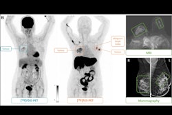
FDG PET/CT also has limitations in certain low-grade and lobular breast cancers, where lesions may show relatively low tracer uptake. Woitek discussed the potential of PET/MRI and targeted radiotracers, including fluorine-18 fluoroestradiol (FES) PET and HER2-targeted PET, to provide information about tumor biology.
Receptor expression can differ between metastatic lesions. Targeted imaging may help identify this heterogeneity across the body and provide additional information for treatment selection.
Woitek also addressed early response assessment using FDG PET and emerging techniques such as hyperpolarized carbon-13 MRI. Their broader clinical use, however, requires further evidence.
When PET and MRI disagree
Dr. Marius E. Mayerhöfer of the Medical University of Vienna in Vienna, Austria, presented cases emphasizing the complementary strengths and limitations of PET and MRI.
In prostate cancer, prostate-specific membrane antigen (PSMA) PET and multiparametric MRI can both detect suspicious disease in the same patient but identify lesions in different locations.
This matters when imaging guides targeted biopsies or treatment planning. Agreement at the patient level does not necessarily translate into agreement on individual lesions.
Mayerhöfer also discussed plasma cell disorders, where CT, FDG PET, and MRI offer different diagnostic advantages. PET/MRI showed the highest sensitivity in the comparative analysis cited during the session.
However, greater sensitivity alone does not establish better clinical outcomes. The value of combining imaging techniques ultimately depends on whether the additional findings improve patient management.