A comprehensive review of 29 studies found no definitive evidence for optimal x-ray beam quality in adult chest radiography despite over two decades of research, highlighting the need for standardized, clinically validated methods to optimize beam quality while reducing radiation exposure.
- Research gap: Despite 20+ years of studies, there is still no consensus on optimal x-ray beam quality for adult chest imaging
- Tube voltage range: Studies most frequently recommended 90-110 kV, but this represented only 43% of reviewed research
- Filtration findings: All 10 studies examining additional filtration recommended its use, primarily aluminum, copper, or combinations
- Digital complication: Digital processing can mask gradual overexposure or dose creep by making different exposures appear similar
- Policy impact: European Union's Euratom Basic Safety Standards require medical exposure optimization, making standardized methods essential
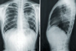
Despite more than two decades of research, there is still no definitive evidence for an optimal x-ray beam quality in adult chest radiography, according to an Irish-led review published in in Physica Medica.
Clare Devery, DipIPEM, a senior medical physicist at St. James’s Hospital and doctoral researcher at Technological University Dublin in Dublin, Ireland, and colleagues examined 29 studies published between 2001 and 2020.
The researchers conducted their literature search in April 2025 using PubMed, Web of Science, and Google Scholar. After removing duplicates, they screened 1,151 records and included 29 studies investigating optimization of posterior-anterior adult chest x-rays.
Wide variation limits firm conclusions
The studies differed considerably in their objectives and methods. Nine focused primarily on reducing radiation dose, seven prioritized improving image quality at a defined dose, and 13 combined both aims. The researchers identified 240 possible combinations of methodological variables, including the source images, detector system, dose metric, image-assessment method, and use of expert readers.
Tube voltages between 90 kV and 110 kV were recommended most frequently, but this range accounted for only 43% of studies. Only 10 studies evaluated additional filtration, although all 10 recommended incorporating some form of filtration, most commonly aluminum, copper, or a combination, into the optimization strategy.
Beam quality reflects the energy and penetration of the x-ray spectrum and is principally influenced by tube voltage and filtration. Higher tube voltage produces a more penetrating beam and may permit lower tube output, while filtration removes low-energy photons that contribute to patient dose without necessarily improving diagnostic information.
Digital imaging complicates optimization
However, the optimal balance also depends on the detector, automatic exposure control, patient size, clinical task, and postprocessing. Digital processing can make images produced with substantially different exposures look similar, potentially concealing gradual overexposure or “dose creep.”
The findings build on earlier coverage of copper filtration as a dose-reduction strategy and warnings that digital cropping can contribute to “collimation creep.” The new review suggests that the larger problem is the absence of a standardized, clinically validated method for determining optimal beam quality.
Findings carry European policy relevance
This has European policy relevance. The European Union’s Euratom Basic Safety Standards Directive requires medical exposures to be optimized while retaining the information needed for diagnosis. Diagnostic reference levels can identify unusually high or low exposures, but they are not dose limits and do not establish whether a protocol provides the best diagnostic performance.
The review also has limitations. It excluded pediatric, film-screen, simulated-image, model-observer, and non-English studies. Most included papers examined beam quality separately from positioning, postprocessing, and clinical indication, while phantom findings may not represent patients of different sizes or the detection of subtle disease.
The authors recommend standardized research methods, transparent reporting of postprocessing, combined evaluation of tube voltage and filtration, and greater involvement of radiologists and reporting radiographers. Implementation would require medical-physics resources, staff training, and equipment-specific validation, but could reduce collective radiation exposure and unwarranted variation between hospitals.
The authors declared no competing interests. Read the full study here.