
Dear AuntMinnieEurope Member,
Prof. Valérie Vilgrain is a towering figure in radiology education. Over the years, she's received many plaudits for her work with the European School of Radiology and the European Society for Gastrointestinal & Abdominal Radiology. She's also an innovator, as shown by her upcoming winter course.
To kick off our coverage of ECR 2025, we asked her to look ahead to this year's congress. Don't miss this eight-minute video interview with Frances Rylands-Monk. Over the coming weeks, we'll bring you further chats with other key players.
In other news, we've posted an article today about an important paper from the researchers behind the SCOT-HEART trial. Radiologist and lead investigator Prof. Michelle Williams speaks about the main findings, new analysis, and the follow-up plans.
Also this week, we posted another thought-provoking column from Dr. Chris Hammond. He's concerned that imaging demand is increasing by about 5% a year in the U.K., and for complex cross-sectional imaging, demand growth was 11% in 2023.
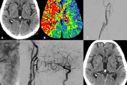
Endovascular thrombectomy is proving valuable in cases of acute extracranial internal carotid artery occlusions, but how does it compare with best medical treatment such as intravenous thrombolysis? German researchers have provided some answers.
Finally, don't forget to check out market expert Amy Thompson's top 5 predictions for imaging IT and AI sectors.
Philip Ward
Editor in Chief
AuntMinnieEurope.com


![Overview of the study design. (A) The fully automated deep learning framework was developed to estimate body composition (BC) (defined as subcutaneous adipose tissue [SAT] in liters; visceral adipose tissue [VAT] in liters; skeletal muscle [SM] in liters; SM fat fraction [SMFF] as a percentage; and intramuscular adipose tissue [IMAT] in deciliters) from MRI. The fully automated framework comprised one model (model 1) to quantify different BC measures (SAT, VAT, SM, SMFF, and IMAT) as three-dimensional (3D) measures from whole-body MRI scans. The second model (model 2) was trained to identify standardized anatomic landmarks along the craniocaudal body axis (z coordinate field), which allowed for subdividing the whole-body measures into different subregions typically examined on clinical routine MRI scans (chest, abdomen, and pelvis). (B) BC was quantified from whole-body MRI in over 66,000 individuals from two large population-based cohort studies, the UK Biobank (UKB) (36,317 individuals) and the German National Cohort (NAKO) (30,291 individuals). Bar graphs show age distribution by sex and cohort. BMI = body mass index. (C) After the performance assessment of the fully automated framework, the change in BC measures, distributions, and profiles across age decades were investigated. Age-, sex-, and height-adjusted body composition reference curves were calculated and made publicly available in a web-based z-score calculator (https://circ-ml.github.io).](https://img.auntminnieeurope.com/mindful/smg/workspaces/default/uploads/2026/05/body-comp.XgAjTfPj1W.jpg?auto=format%2Ccompress&dpr=2&fit=crop&h=167&q=70&w=250)


