
Dear AuntMinnieEurope Member,
News broke on Wednesday in the U.K. media about an important medicolegal case.
A coroner found a radiologist's failure to identify a slipped gastric band was a critical factor in the death of a 44-year-old woman. He issued a Prevent Future Deaths report to National Health Service (NHS) England and the U.K. Royal College of Radiologists (RCR), and this has prompted the RCR to assess how it can boost awareness of slipped gastric bands.
In other U.K. news, an AI action plan was launched on Monday. The 26-page report has received much praise, but implementing the 50 recommendations made by AI entrepreneur Matt Clifford represents a huge challenge, some observers believe. If you missed our coverage, you can read it here.
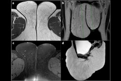
Thankfully, vulvar pathologies are rare -- but because of their rarity, many radiologists struggle to diagnose some conditions correctly. This prompted a research team from Vienna and Rome to produce an authoritative article on MRI of the vulva. The authors kindly agreed to share four sets of clinical images with us.
Also this week, we posted an entertaining new column from Dr. Paul McCoubrie. He's a fine writer with bold and original views, and he's always worth listening to.
Finally, Dutch investigators have found that a deep-learning model based on MRI and clinical parameters appears to be effective for predicting the risk of prostate cancer progression.
Looking ahead to next week, we'll be launching our build-up to ECR 2025. Associate Editor Frances Rylands-Monk has recorded video interviews with some of the key players, so watch this space.
Philip Ward
Editor in Chief
AuntMinnieEurope.com


![Overview of the study design. (A) The fully automated deep learning framework was developed to estimate body composition (BC) (defined as subcutaneous adipose tissue [SAT] in liters; visceral adipose tissue [VAT] in liters; skeletal muscle [SM] in liters; SM fat fraction [SMFF] as a percentage; and intramuscular adipose tissue [IMAT] in deciliters) from MRI. The fully automated framework comprised one model (model 1) to quantify different BC measures (SAT, VAT, SM, SMFF, and IMAT) as three-dimensional (3D) measures from whole-body MRI scans. The second model (model 2) was trained to identify standardized anatomic landmarks along the craniocaudal body axis (z coordinate field), which allowed for subdividing the whole-body measures into different subregions typically examined on clinical routine MRI scans (chest, abdomen, and pelvis). (B) BC was quantified from whole-body MRI in over 66,000 individuals from two large population-based cohort studies, the UK Biobank (UKB) (36,317 individuals) and the German National Cohort (NAKO) (30,291 individuals). Bar graphs show age distribution by sex and cohort. BMI = body mass index. (C) After the performance assessment of the fully automated framework, the change in BC measures, distributions, and profiles across age decades were investigated. Age-, sex-, and height-adjusted body composition reference curves were calculated and made publicly available in a web-based z-score calculator (https://circ-ml.github.io).](https://img.auntminnieeurope.com/mindful/smg/workspaces/default/uploads/2026/05/body-comp.XgAjTfPj1W.jpg?auto=format%2Ccompress&dpr=2&fit=crop&h=167&q=70&w=250)


