New research from LMU University Hospital in Munich has found that PET imaging helps clinicians predict survival outcomes for patients with brain tumors. The study findings were published on 3 August in the Journal of Nuclear Medicine.
A group led by Dr. Nathalie Albert, who is a member of the European Association of Nuclear Medicine's neuroimaging committee, linked translocator protein (TSPO)-PET imaging and shorter survival in patients with newly diagnosed glioblastoma undergoing radiotherapy.
"TSPO-PET seems to add prognostic insights beyond established clinical parameters and might serve as an informative tool as clinicians make survival predictions," the researchers wrote.
Glioblastoma is the most frequent malignant primary brain tumor in adults, and diagnosis is associated with a short life expectancy. TSPO is a protein expressed in glioblastoma tumors. PET radiotracers designed to bind to TSPO can reveal tumor characteristics, yet studies showing the clinical benefit of the approach remain scarce, the authors explained.
To that end, the group investigated associations between TSPO-PET imaging using F-18 GE-180 radiotracer with survival outcomes in 45 patients with histologically proven isocitrate dehydrogenase (IDH) wild-type glioblastoma prior to undergoing radiotherapy treatment.
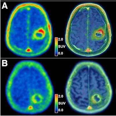
 (A) A 76-year-old male patient with left precentral IDH wild-type glioblastoma before hypofractionated radiotherapy and temozolomide chemotherapy. Tumoral TSPO radioligand uptake was high (SUVmax, 2.4), and survival was short (OS, 4.5 months). (B) A 71-year-old female patient with left postcentral parietal IDH wild-type glioblastoma before hypofractionated radiotherapy and temozolomide chemotherapy. Tumoral TSPO radioligand uptake was low (SUVmax, 1.8), and survival was long (OS, 25.8 months). Image and caption courtesy of the Journal of Nuclear Medicine (CC BY 4.0).
The patients' median progression-free survival was 8.1 months and overall survival was 10.8 months. At the last treatment follow-up, 43 of 45 patients (95.6%) had experienced tumor progression and 40 of 45 patients (88.9%) had died.
According to the findings, tumoral uptake of F-18 GE-180 on the PET imaging was associated with overall survival. Specifically, a high maximum standard uptake value (SUVmax) was related to significantly shorter overall survival: 8.3 months versus 17.8 months, the researchers found.
"Compared with patients with low tumoral uptake on TSPO-PET, patients with SUVmax of more than 2.2 had a significantly higher risk for death, with a hazard ratio of 2.2 in the multivariate analysis," the group wrote.
Ultimately, successful treatment options remain limited for patients with glioblastoma, and there is a need to explore new targets for both diagnostics and therapy in these patients. This study suggests that TSPO-PET imaging during radiotherapy is a promising tool for assessing treatment response, the authors noted.
"High tumoral uptake in TSPO-PET before radiotherapy is associated with significantly shorter survival within the homogeneous group of molecularly defined, newly diagnosed IDH wild-type glioblastoma," the group concluded.
The full study can be found here.